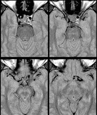
Intracranial dolichoectasias
Topic: Medicine
 From HandWiki - Reading time: 2 min
| Intracranial dolichoectasias | |
|---|---|
 |
The term dolichoectasia means elongation and distension. It is used to characterize arteries throughout the human body which have shown significant deterioration of their tunica intima (and occasionally the tunica media), weakening the vessel walls and causing the artery to elongate and distend.
Signs and symptoms
VBD
- Hemifacial spasm
- Paresis
- Trigeminal neuralgia
ICD
- Progressive visual field defect
Cause
Most commonly caused by hypertension, continued stress on the walls of the artery will degrade the vessel wall by damaging and loosening the collagen and elastin meshwork which comprises the intima. Similarly, hypercholesterolemia or hyperlipidemia can also provide sufficient trauma to the vessel wall resulting in dolichoectasia. As the arrangement of connective tissue is disturbed, the vessel wall is no longer able to hold its original conformation and begins to unravel due to the continued hypertension. High blood pressure mold and force the artery to now take on an elongated, tortuous course to better withstand the higher pressures.
Pathophysiology
Most commonly affected is the Vertebral Basilar Artery (Vertebral Basilar Dolichoectasia or Vertebrobasillar Dolichoectasia). The Internal Carotid Artery is also at high risk to be affected. Patients with Autosomal Dominant Polycystic Kidney Disease (ADPKD) are more likely to be subject to dolichoectasias. Dolichoectasias are most common in elderly males.[1]
In cases involving the basilar artery (VBD), the pathogenesis arises from direct compression of different cranial nerves.
Additionally, ischemic effects on the brain stem and cerebellar hemispheres as well as symptoms related to hydrocephalus are common. Direct cranial nerve compression can lead to isolated cranial nerve dysfunction, usually associated with a normal-sized basilar artery that is tortuous and elongated. Cranial nerve dysfunction most commonly involves the VII cranial nerve and the V cranial nerve. Multiple cranial nerve dysfunction is far more likely to occur if there is dilation (ectasia) associated with a tortuous and elongated basilar artery. Cranial nerves affected in descending order of frequency include: VII, V, III, VIII, and VI.
Internal Carotid Artery dolichoectasia is particularly interesting because the artery normally already contains one hairpin turn. Seen in an MRI as two individual arteries at this hairpin, a carotid artery dolichoectasia can progress so far as to produce a second hairpin turn and appear as three individual arteries on an MRI. In the case of a dolichoectasia of the Internal Carotid Artery (ICD), the pathogenesis is primarily related to compression of the Optic Nerves at the Optic Chiasma (see Fig. 1 and 2).
Diagnosis
Treatment
A combination of lifestyle modifications and medications can be used for the treatment of dolichoectasias.
- Antihypertensive medications such as Thiazides, Beta Blocker, ACE Inhibitor
- Trental or other Pentoxifylline drugs
- Dietary changes
- Weight loss
- Regular exercise
References
- ↑ Yu, YL; Moseley, IF; Pullicino, P; McDonald, WI (1982). "The clinical picture of ectasia of the intracerebral arteries.". J Neurol Neurosurg Psychiatry 45 (1): 29–36. doi:10.1136/jnnp.45.1.29. ISSN 0022-3050.
- Vertebrobasilar Dolichoectasia, rad.usuhs.mil
- Dolichoectasia of Internal Carotid Artery[|permanent dead link|dead link}}], medcyclopaedia.com (archived from the original)
- Relation between ADPKD and Dolichoectasia
 KSF
KSF
