Long ciliary nerves
Topic: Medicine
 From HandWiki - Reading time: 2 min
| Long ciliary nerves | |
|---|---|
 Nerves of the orbit, and the ciliary ganglion. Side view. | |
| Details | |
| From | Nasociliary nerve |
| Innervates | Cornea, iris, and ciliary body |
| Fiber type | "Somatosensory" (via V1 Lacrimal), and "Sympathetic" (via V2 Zygomatic) |
| Identifiers | |
| Latin | nervi ciliares longi |
| Anatomical terms of neuroanatomy | |
The long ciliary nerves are two-three sensory nerves that arise from the nasociliary nerve (itself a branch of the ophthalmic branch (CN V1) of the trigeminal nerve (CN V)).[1] They pass forward within the orbit, passing toward the eyeball alongside the optic nerve (CN II).[2] They enter the eyeball to provide sensory innervation to the cornea, iris, and ciliary body.[3] They also provide sympathetic visceral motor innervation to the dilator pupillae muscle, which is responsible for dilation of the pupil.[4] The long ciliary nerves are clinically relevant in conditions affecting corneal sensitivity, pupillary responses, and surgical procedures involving the eye.[5]
Anatomy
Origin
The long ciliary nerves branch from the nasociliary nerve as it crosses the optic nerve (CN II).[1]
Course
Accompanied by the short ciliary nerves, the long ciliary nerves pierce and enter[1] the posterior part of the sclera near where it is entered by the optic nerve, then run anterior-ward between the sclera and the choroid.[1]
Function
The long ciliary nerves are distributed to the ciliary body, iris, and cornea.[1]
Sensory
The long ciliary nerves provide sensory innervation to the eyeball, including the cornea.[6]
Sympathetic
See also
- Short ciliary nerves
Additional images
-
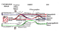 Pathways in the ciliary ganglion.

References
- ↑ 1.0 1.1 1.2 1.3 1.4 Standring, Susan (2020). Gray's Anatomy: The Anatomical Basis of Clinical Practice (42nd ed.). New York. pp. 783. ISBN 978-0-7020-7707-4. OCLC 1201341621.
- ↑ Ansari, Mohammad Wakeel; Nadeem, Ahmed (2016). "Atlas of Ocular Anatomy". Ocular Anatomy. doi:10.1007/978-3-319-42781-2. https://link.springer.com/book/10.1007/978-3-319-42781-2?error=cookies_not_supported&code=988742a0-3716-4576-89ca-a88746e5dca8.
- ↑ Levin, Leonard A.; Kaufman, Paul L.; Hartnett, Mary Elizabeth (2024). Adler's physiology of the eye (12th ed.). Chantilly: Elsevier. ISBN 978-0-323-83407-0.
- ↑ Wu, Feipeng; Zhao, Yin; Zhang, Hong (2022-01-14). "Ocular Autonomic Nervous System: An Update from Anatomy to Physiological Functions" (in en). Vision 6 (1): 6. doi:10.3390/vision6010006. ISSN 2411-5150. PMID 35076641. PMC 8788436. https://www.mdpi.com/2411-5150/6/1/6.
- ↑ Lum, Edward; Corbett, Melanie C.; Murphy, Paul J. (July 2019). "Corneal Sensitivity After Ocular Surgery" (in en). Eye & Contact Lens: Science & Clinical Practice 45 (4): 226–237. doi:10.1097/ICL.0000000000000543. ISSN 1542-2321. https://journals.lww.com/00140068-201907000-00003.
- ↑ Yang, Alina Y.; Chow, Jessica; Liu, Ji (March 2018). "Corneal Innervation and Sensation: The Eye and Beyond". The Yale Journal of Biology and Medicine 91 (1): 13–21. ISSN 1551-4056. PMID 29599653. PMC 5872636. https://pubmed.ncbi.nlm.nih.gov/29599653.
This article incorporates text in the public domain from the 20th edition of Gray's Anatomy (1918)
External links
 |  |
 EncycloReader
is supported by the
EncycloReader
is supported by the  KSF
KSF