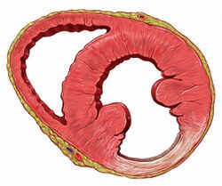
Myocardial scarring
Topic: Medicine
 From HandWiki - Reading time: 8 min

Myocardial scarring is the accumulation of fibrous tissue resulting after some form of trauma to the cardiac tissue.[1][2] Fibrosis is the formation of excess tissue in replacement of necrotic or extensively damaged tissue. Fibrosis in the heart is often hard to detect because fibromas, scar tissue or small tumors formed in one cell line, are often formed.[3] Because they are so small, they can be hard to detect by methods such as magnetic resonance imaging.[1] A cell line is a path of fibrosis that follow only a line of cells.
Causes
Myocardial infarction
A myocardial infarction, also known as a heart attack, often result in the formation of fibrosis.[2] A myocardial infarction is an ischemic event, or a restriction of blood flow to body tissue, such as by atherothrombosis.[4] Without blood flow to the myocardium, it is deprived of oxygen, causing tissue death and irreversible damage.[5] The tissue destroyed by the infarction is replaced with non-functioning fibrosis, restoring some of the structural integrity of the organ but resulting in impaired myocardial function.[6]
Coronary heart disease
Coronary heart disease, also known as coronary artery disease, is one of the most common causes of myocardial damage, affecting over three million people in the United States.[7] In coronary heart disease, the coronary arteries narrow due to the buildup of atheroma or fatty deposits on the vessel walls. The atheroma causes the blood flow of the arteries to be restricted.[6] By restricting the blood flow, the tissue is still receiving some oxygen, but not enough to sustain the tissue over time.[5] The accumulation of the fibrotic tissue is much slower in coronary heart disease compared to an infarction because the tissue is still receiving some oxygen.[6]
Birth defect repairs
Another form of myocardial scarring results from surgical repairs.[2] Surgical repairs are often necessary for a person born with a congenital defect of the heart.[8] While surgical laparoscopy still leaves myocardial scarring, the trauma seems to be less damaging then naturally occurring scarring.[2]
Excessive exercise
Excessive exercise, particularly in endurance athletes, has been linked to myocardial scarring, a condition where fibrous tissue in the heart muscle forms. Intense, prolonged physical activity can lead to repetitive stress on the myocardium, causing micro-injuries that may trigger inflammation and subsequent scarring. This is especially prevalent in individuals engaging in extreme endurance sports, such as marathons or ultra-marathons, very long bicycle racing, where sustained high cardiac output can strain the heart. Studies have shown that some athletes develop focal myocardial fibrosis, detectable via cardiac imaging (cardiac MRI, etc), which may increase the risk of arrhythmias or reduced cardiac function over time. While exercise is generally beneficial for heart health, excessive and prolonged exertion without adequate recovery may contribute to pathological changes in susceptible individuals - genetic tendency toward cardiac fibrosis, undiagnosed heart issues, or a reduced ability to recover from exercise-induced inflammation or micro-injuries.[9][10][11][12]
Hypertension
Chronic hypertension can lead to myocardial scarring by imposing persistent high pressure on heart walls. This stress causes left ventricular hypertrophy, increasing oxygen demand and potentially leading to micro-ischemia. Over time, these changes may result in fibrosis as the heart attempts to repair damaged tissue, impairing cardiac function.[13]
Cardiomyopathies
Cardiomyopathies, such as dilated or hypertrophic cardiomyopathy, are associated with myocardial scarring. These conditions alter heart muscle structure and function. Scarring may contribute to heart failure or arrhythmias.[14]
Myocarditis
Myocarditis, inflammation of the heart muscle often caused by viral infections, can result in myocardial scarring. The inflammatory response damages cardiac tissue, triggering repair processes that replace necrotic cells with fibrous tissue. This scarring may increase the risk of arrhythmias.[15][16]
Cardiac toxins
Exposure to cardiotoxic substances, such as chemotherapy drugs (e.g., anthracyclines), alcohol, or recreational drugs like cocaine, can cause myocardial scarring due to free radical damage, strand breakage of DNA in cardiomyocytes.[17] These toxins induce direct damage to heart muscle cells, leading to inflammation and fibrosis as the body attempts to repair the injured tissue.[18][19][20]
Aging
Aging is a natural contributor to myocardial scarring. Over time, cumulative stress from oxidative damage, low-grade inflammation, and microvascular changes can lead to gradual fibrosis in the heart. This age-related scarring may reduce cardiac elasticity and contribute to diastolic dysfunction in older individuals.[21][22]
Formation
Immediately after damage to the myocardium occurs, the damaged tissue becomes inflamed. Inflammation is the accumulations of neutrophils, macrophages, and lymphocytes at the site of the trauma.[23][24] In addition, "inflammatory cells upregulate the release of a myriad of signaling cytokines, growth factors, and hormones including transforming growth factor β, interleukins 1, 2, 6, and 10, tumor necrosis factor α, interferon γ, chemokines of the CC and CXC families, angiotensin II, norepinephrine, endothelin, natriuretic peptides, and platelet-derived growth factors".[24] Both the necrotic cells and the inflamed myocardium secrete and activate matrix metalloproteinase. Metalloproteinase aids in the destruction and reabsorption of necrotic tissue. After several days, collagen accumulation at the site of injury begins to occur.[24] As part of the extracellular matrix, granulated tissue consisting of fibrin, fibronectin, laminin, and glycosaminoglycan is suspended in a collagen base.[24] The extracellular matrix acts as scaffolding for the fibrillar collagen to form. The fibrillar collagen is the main constitute of what will become the scar tissue.[24]
References
- ↑ 1.0 1.1 Guler, Gamze Babur (2011). "Myocardial Fibrosis Detected by Cardiac Magnetic Resonance Imaging in Heart Failure: Impact on Remodeling, Diastolic Function and BNP Levels". Anatolian Journal of Cardiology 11 (1): 71–76. doi:10.5152/akd.2011.013. PMID 21220243.
- ↑ 2.0 2.1 2.2 2.3 Fomovsky, Gregory M. (2010). "Evolution of Scar Structure, Mechanics, and Ventricular Function after Myocardial Infarction in the Rat". American Journal of Physiology. Heart and Circulatory Physiology 298 (1): 1–12. doi:10.1152/ajpheart.00495.2009. PMID 19897714.
- ↑ (in en).
- ↑ Katz, Monica Y. (2014). "Three-Dimensional Myocardial Scarring along Myofibers after Coronary Ischemia-Reperfusion Revealed by Computerized Images of Histological Assays". Physiological Reports 2 (7): 1–3. doi:10.14814/phy2.12072. PMID 25347856.
- ↑ 5.0 5.1 (in en).
- ↑ 6.0 6.1 6.2 Liang, Cuiping (2019). "Influence of the Distribution of Fibrosis within an Area of Myocardial Infarction on Wave Propagation in Ventricular Tissue". Scientific Reports 15 (1): 1–24. doi:10.1038/s41598-019-50478-5. PMID 31578428. Bibcode: 2019NatSR...914151L.
- ↑ (in en).
- ↑ CDC (2019-11-22). "What are Congenital Heart Defects? | CDC" (in en-us). https://www.cdc.gov/ncbddd/heartdefects/facts.html.
- ↑ Patil, Harshal R.; O'Keefe, James H.; Lavie, Carl J.; Magalski, Anthony; Vogel, Robert A.; McCullough, Peter A. (2012). "Cardiovascular damage resulting from chronic excessive endurance exercise". Missouri Medicine 109 (4): 312–321. ISSN 0026-6620. PMID 22953596.
- ↑ "Heart Risks Associated With Extreme Exercise" (in en). https://health.clevelandclinic.org/can-too-much-extreme-exercise-damage-your-heart.
- ↑ O'Keefe, James H.; Patil, Harshal R.; Lavie, Carl J.; Magalski, Anthony; Vogel, Robert A.; McCullough, Peter A. (June 2012). "Potential adverse cardiovascular effects from excessive endurance exercise". Mayo Clinic Proceedings 87 (6): 587–595. doi:10.1016/j.mayocp.2012.04.005. ISSN 1942-5546. PMID 22677079.
- ↑ "New research to better understand how heart scarring impacts veteran athletes" (in en). https://www.bhf.org.uk/what-we-do/news-from-the-bhf/news-archive/2022/may/new-research-to-better-understand-how-heart-scarring-impacts-veteran-athletes.
- ↑ Saheera, Sherin; Krishnamurthy, Prasanna (2020). "Cardiovascular Changes Associated with Hypertensive Heart Disease and Aging". Cell Transplantation 29. doi:10.1177/0963689720920830. ISSN 1555-3892. PMID 32393064.
- ↑ El Hadi, Hamza; Freund, Anne; Desch, Steffen; Thiele, Holger; Majunke, Nicolas (2023-02-11). "Hypertrophic, Dilated, and Arrhythmogenic Cardiomyopathy: Where Are We?". Biomedicines 11 (2): 524. doi:10.3390/biomedicines11020524. ISSN 2227-9059. PMID 36831060.
- ↑ Kang, Michael; Chippa, Venu; An, Jason (2025), "Viral Myocarditis", StatPearls (Treasure Island (FL): StatPearls Publishing), PMID 29083732, https://www.ncbi.nlm.nih.gov/books/NBK459259/, retrieved 2025-05-29
- ↑ Shams, Pirbhat; Collier, Sara A. (2025), "Acute Myocarditis", StatPearls (Treasure Island (FL): StatPearls Publishing), PMID 28722877, https://www.ncbi.nlm.nih.gov/books/NBK441847/, retrieved 2025-05-29
- ↑ Chong, Esther G.; Lee, Eric H.; Sail, Reena; Denham, Laura; Nagaraj, Gayathri; Hsueh, Chung-Tsen (2021-01-26). "Anthracycline-induced cardiotoxicity: A case report and review of literature". World Journal of Cardiology 13 (1): 28–37. doi:10.4330/wjc.v13.i1.28. ISSN 1949-8462. PMID 33552401.
- ↑ Morelli, Marco Bruno; Bongiovanni, Chiara; Da Pra, Silvia; Miano, Carmen; Sacchi, Francesca; Lauriola, Mattia; D'Uva, Gabriele (2022). "Cardiotoxicity of Anticancer Drugs: Molecular Mechanisms and Strategies for Cardioprotection". Frontiers in Cardiovascular Medicine 9. doi:10.3389/fcvm.2022.847012. ISSN 2297-055X. PMID 35497981.
- ↑ Mabudian, Leila; Jordan, Jennifer H.; Bottinor, Wendy; Hundley, W. Gregory (2022-09-27). "Cardiac MRI assessment of anthracycline-induced cardiotoxicity" (in English). Frontiers in Cardiovascular Medicine 9. doi:10.3389/fcvm.2022.903719. ISSN 2297-055X. PMID 36237899.
- ↑ Adão, Rui; de Keulenaer, Gilles; Leite-Moreira, Adelino; Brás-Silva, Carmen (2013-05-01). "Cardiotoxicity associated with cancer therapy: Pathophysiology and prevention" (in pt). Revista Portuguesa de Cardiologia 32 (5): 395–409. doi:10.1016/j.repce.2012.11.019. ISSN 0870-2551. http://www.revportcardiol.org/pt-cardiotoxicidade-associada-a-terapeutica-oncologica-mecanismos-fisiopatologicos-e-articulo-S2174204913000895.
- ↑ Horn, Margaux A.; Trafford, Andrew W. (April 2016). "Aging and the cardiac collagen matrix: Novel mediators of fibrotic remodelling". Journal of Molecular and Cellular Cardiology 93: 175–185. doi:10.1016/j.yjmcc.2015.11.005. ISSN 1095-8584. PMID 26578393.
- ↑ Wood, Colby; Salter, Wm Zachary; Garcia, Isaiah; Nguyen, Michelle; Rios, Andres; Oropeza, Jacqui; Ugwa, Destiny; Mukherjee, Upasana et al. (2025-05-13). "Age-associated changes in the heart: implications for COVID-19 therapies" (in en). Aging 17. doi:10.18632/aging.206251. ISSN 1945-4589. PMID 40372276. PMC 12151517. https://www.aging-us.com/article/206251/text.
- ↑ Radauceanu, Anca (2007). "Residual Stress Ischemia Is Associated with Blood Markers of Myocardial Structural Remodeling". European Journal of Heart Failure 9 (4): 370–376. doi:10.1016/j.ejheart.2006.09.010. PMID 17140850.
- ↑ 24.0 24.1 24.2 24.3 24.4 Richardson, William J.; Clarke, Samantha A.; Quinn, T. Alexander; Holmes, Jeffrey W. (2015-09-20). "Physiological Implications of Myocardial Scar Structure". Comprehensive Physiology 5 (4): 1877–1909. doi:10.1002/cphy.c140067. ISSN 2040-4603. PMID 26426470.
 |
 KSF
KSF