Antigen-presenting cell
 From Wikidoc - Reading time: 4 min

Editor-In-Chief: C. Michael Gibson, M.S., M.D. [1]
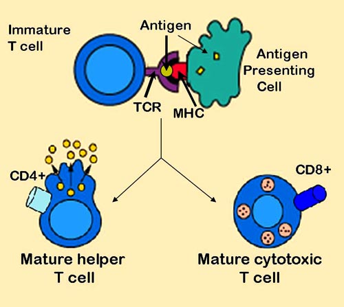
Template:Seealso An antigen-presenting cell (APC) or accessory cell is a cell that displays foreign antigen complexed with MHC on its surface. T-cells may recognize this complex using their T-cell receptor (TCR).
Types[edit | edit source]
APCs fall into two categories: professional or non-professional.
Although almost every cell in the body is an APC, since it can present antigen to CD8+ T cells via MHC class I molecules, the term is often limited to those specialized cells that can prime T cells (i.e., activate a T cell that has not been exposed to antigen, termed a naive T cell). These cells, in general, express MHC class II as well as MHC class I molecules, and can stimulate CD4+ ("helper") cells as well as CD8+ ("cytotoxic") T cells.
To help distinguish between the two types of APCs, those that express MHC class II molecules are often called professional antigen-presenting cells.
Professional APCs[edit | edit source]
These professional APCs are very efficient at internalizing antigen, either by phagocytosis or by receptor-mediated endocytosis, and then displaying a fragment of the antigen, bound to a class II MHC molecule, on their membrane. The T cell recognizes and interacts with the antigen-class II MHC molecule complex on the membrane of the antigen-presenting cell. An additional co-stimulatory signal is then produced by the antigen-presenting cell, leading to activation of the T cell.
There are three main types of professional antigen-presenting cells:
- Dendritic cells, which have the broadest range of antigen presentation, and are probably the most important APC. Activated DCs are especially potent TH cell activators because, as part of their composition, they express co-stimulatory molecules such as B7.
- Macrophages
- B-cells, which express antibody, can very efficiently present the antigen to which their antibody is directed, but are inefficient APC for most other antigens.
As well, there are specialized cells in particular organs (e.g., microglia in the brain, Kupffer cells in the liver) derived from macrophages that are also effective APCs.
Non-professional[edit | edit source]
A non-professional APC does not constitutively express the Major histocompatibility complex proteins required for interaction with naive T cells; these are expressed only upon stimulation of the non-professional APC by certain cytokines such as IFN-γ. Non-professional APCs include:
- Fibroblasts (skin)
- Thymic epithelial cells
- Thyroid epithelial cells
- Glial cells (brain)
- Pancreatic beta cells
- Vascular endothelial cells
Interaction with T cells[edit | edit source]
After dendritic cells or macrophages swallow pathogens, they usually migrate to the lymph nodes, where most T cells are. They do this by chemotaxis: Chemokines that flow in the blood and lymph vessels "draw" the APCs to the lymph nodes. During the migration, DCs undergo a process of maturation; in essence, they lose most of their ability to further swallow pathogens, and they develop an increased ability to communicate with T cells. Enzymes within the cell digest the swallowed pathogen into smaller pieces containing epitopes, which are then presented to T cells using MHC.
Recent research indicates that only certain epitopes of a pathogen are presented because they are immunodominant, possibly as a function of their binding affinity to the MHC. The stronger binding affinity allows the complex to remain kinetically stable long enough to be recognized by T cells.
External links[edit | edit source]
- Antigen-Presenting+Cells at the US National Library of Medicine Medical Subject Headings (MeSH)
de:Antigenpräsentierende Zelle
ko:항원전달세포
it:Cellula APC
sv:Antigenpresenterande cell
 KSF
KSF