Cirrhosis CT
 From Wikidoc - Reading time: 4 min
|
Cirrhosis Microchapters |
|
Diagnosis |
|---|
|
Treatment |
|
Case studies |
|
Cirrhosis CT On the Web |
|
American Roentgen Ray Society Images of Cirrhosis CT |
Editor-In-Chief: C. Michael Gibson, M.S., M.D. [1]; Associate Editor(s)-in-Chief: Sudarshana Datta, MD [2]
Overview[edit | edit source]
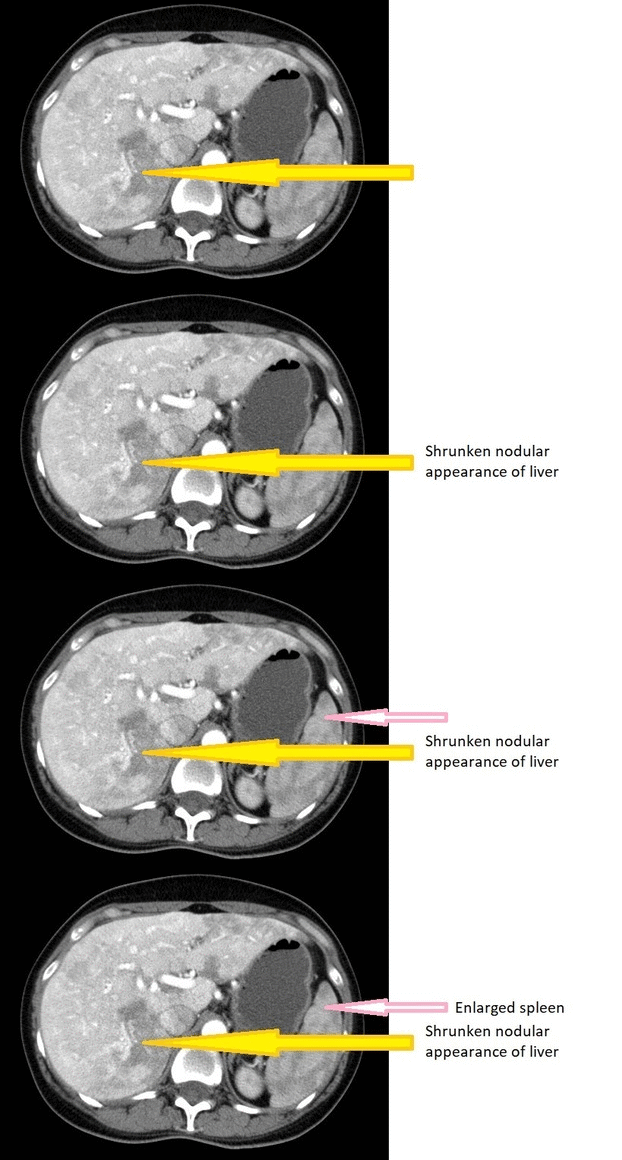
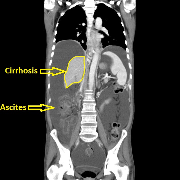
Although CT scans are not routinely used in evaluation and diagnosis of cirrhosis, it may show the presence of lobar atrophic and hypertrophic changes in the liver, ascites and varices. CT scans also visualize the presence of tumors, blocked bile ducts and help evaluate the size of the liver.
CT[edit | edit source]
- Computed tomography is not routinely used in the diagnosis and evaluation of cirrhosis.
- Computed tomography (CT) scanning complements ultrasound imaging.
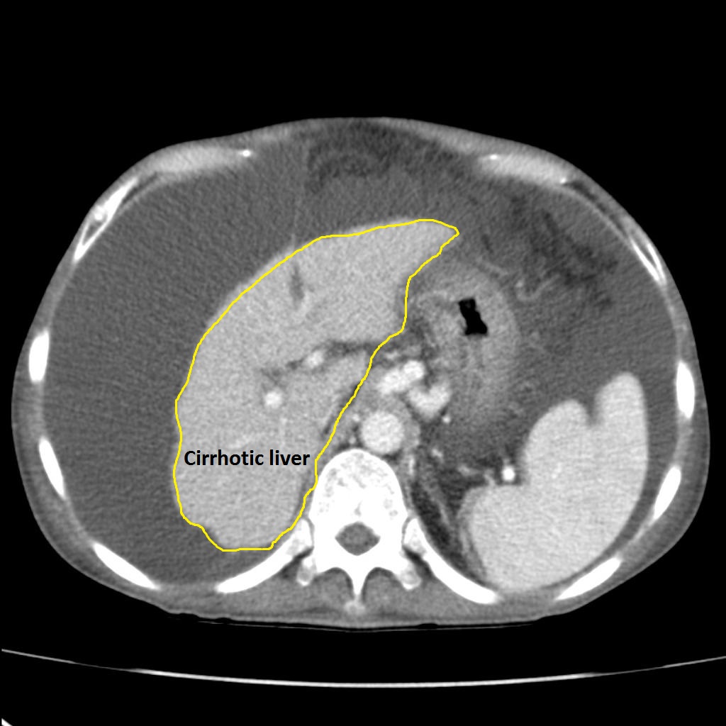
- CT scan is poor at detecting morphologic changes associated with early cirrhosis, but may accurately demonstrate nodularity and lobar atrophic and hypertrophic changes, ascites and varices in advanced disease.
- CT findings may suggest the presence of cirrhosis, but is not diagnostic.
- CT portal phase imaging may be used in the assesment of patency of the portal vein.[1]
- CT may be indicative of underlying etiology due to its classical appearances in some diseases:
- Budd-Chiari syndrome: hypertrophied caudate lobe
- Haemochromatosis: excess iron deposition leads to a dramatic increase in hepatic density
- CT scan in patients with cirrhosis may be used to detect:
- Hepatic nodularity
- Atrophy of the right lobe
- Hypertrophy of the caudate or left lobes
- Ascites
- Varices
- Liver size
- Blocked bile ducts
- Blood flow through the liver
- Tumors
- Side effects of CT scans:
- Exposure to contrast and radiation
CT Images[edit | edit source]

Source: Wikimedia commons [2]
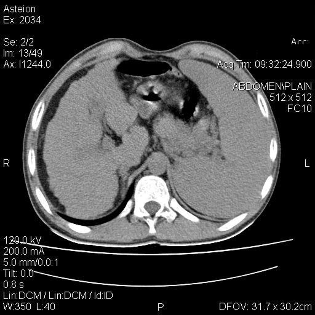
- Abdominal CT scan may be helpful in the diagnosis of portal hypertension. Findings on CT scan suggestive of portal hypertension include:[3][4][5][6]
- Cirrhotic liver, as shrinkage and atrophy in liver
- Re-canalized umbilical vein--pathognomonic
- Dilated portal vein and/or splanchnic veins
- Esophageal varices
- Collaterals in any abdominal organ
- Splenomegaly
- Ascites
Portal hypertension
 |
 |
 |
 |
Recanalized Umbilical Vein
 |
 |
 |
 |
 |
 |
References[edit | edit source]
- ↑ "Cirrhosis and Chronic Liver Failure: Part I. Diagnosis and Evaluation - September 1, 2006 - American Family Physician". Retrieved 2012-09-07.
- ↑ "File:Morbus-Osler-CT-Leber-ax-012.jpg - Wikimedia Commons". External link in
|title=(help) - ↑ Procopet, Bogdan; Berzigotti, Annalisa (2017). "Diagnosis of cirrhosis and portal hypertension: imaging, non-invasive markers of fibrosis and liver biopsy". Gastroenterology Report. 5 (2): 79–89. doi:10.1093/gastro/gox012. ISSN 2052-0034.
- ↑ Aagaard, J; Jensen, LI; Sorensen, TI; Christensen, U; Burcharth, F (1982). "Recanalized umbilical vein in portal hypertension". American Journal of Roentgenology. 139 (6): 1107–1110. doi:10.2214/ajr.139.6.1107. ISSN 0361-803X.
- ↑ Cho, K C; Patel, Y D; Wachsberg, R H; Seeff, J (1995). "Varices in portal hypertension: evaluation with CT". RadioGraphics. 15 (3): 609–622. doi:10.1148/radiographics.15.3.7624566. ISSN 0271-5333.
- ↑ Bandali, Murad Feroz; Mirakhur, Anirudh; Lee, Edward Wolfgang; Ferris, Mollie Clarke; Sadler, David James; Gray, Robin Ritchie; Wong, Jason Kam (2017). "Portal hypertension: Imaging of portosystemic collateral pathways and associated image-guided therapy". World Journal of Gastroenterology. 23 (10): 1735. doi:10.3748/wjg.v23.i10.1735. ISSN 1007-9327.
- ↑ From the case <"https://radiopaedia.org/cases/23057">rID: 23057
- ↑ From the case <"https://radiopaedia.org/cases/23057">rID: 23057
- ↑ From the case <"https://radiopaedia.org/cases/5379">rID: 5379
- ↑ From the case <"https://radiopaedia.org/cases/15852">rID: 15852
- ↑ 11.0 11.1 11.2 11.3 11.4 11.5 Radiopaedia.org. From the case <"https://radiopaedia.org/cases/11810">rID: 11810
Licensed under CC BY-SA 3.0 | Source: https://www.wikidoc.org/index.php/Cirrhosis_CT19 views | Status: cached on October 20 2025 18:23:38↧ Download this article as ZWI file
 KSF
KSF