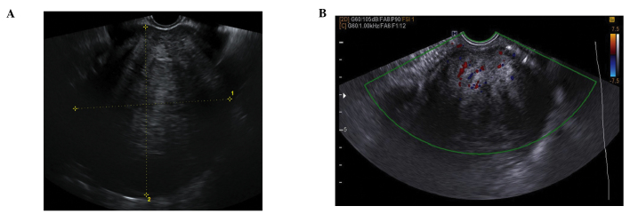
Fibroma ultrasound
 From Wikidoc - Reading time: 3 min
|
Fibroma Microchapters |
|
Diagnosis |
|---|
|
Treatment |
|
Case Studies |
|
Fibroma ultrasound On the Web |
|
American Roentgen Ray Society Images of Fibroma ultrasound |
Editor-In-Chief: C. Michael Gibson, M.S., M.D. [1]; Associate Editor(s)-in-Chief: Maneesha Nandimandalam, M.B.B.S.[2], Simrat Sarai, M.D. [3]
Overview[edit | edit source]
Ultrasound may be helpful in the diagnosis of fibroma. Findings on ultrasound suggestive of fibroma include solid, hypoechoic masses with ultrasound beam attenuation.
Ultrasound[edit | edit source]
On ultrasound, fibromas most commonly manifest as solid, hypoechoic masses with ultrasound beam attenuation.
Ovarian Fibroma[edit | edit source]
On ultrasound, ovarian fibroma most commonly manifest as solid, hypoechoic masses with ultrasound beam attenuation. As such, they may appear similar to a pedunculated subserosal uterine fibroid. However, the sonographic appearance can be variable and some tumors can rarely have cystic components.[1][2][3][4]


Uterine Fibroma[edit | edit source]
- Uncomplicated leiomyomas are usually hypoechoic, but can be isoechoic, or even hyperechoic compared to normal myometrium
- Calcification is seen as echogenic foci with shadowing
- Cystic areas of necrosis or degeneration may be seen
References[edit | edit source]
- ↑ Chen, Hui; Liu, Yan; Shen, Li-fei; Jiang, Mei-jiao; Yang, Zhi-fang; Fang, Guo-ping (2016). "Ovarian thecoma-fibroma groups: clinical and sonographic features with pathological comparison". Journal of Ovarian Research. 9 (1). doi:10.1186/s13048-016-0291-2. ISSN 1757-2215.
- ↑ Liu, Yan; Zhang, Hui; Li, Xiaoqian; Qi, Guiqin (2016). "Combined Application of Ultrasound and CT Increased Diagnostic Value in Female Patients with Pelvic Masses". Computational and Mathematical Methods in Medicine. 2016: 1–5. doi:10.1155/2016/6146901. ISSN 1748-670X.
- ↑ Sayasneh, Ahmad; Ekechi, Christine; Ferrara, Laura; Kaijser, Jeroen; Stalder, Catriona; Sur, Shyamaly; Timmerman, Dirk; Bourne, Tom (2015). "The characteristic ultrasound features of specific types of ovarian pathology (Review)". International Journal of Oncology. 46 (2): 445–458. doi:10.3892/ijo.2014.2764. ISSN 1019-6439.
- ↑ Sconfienza, L.M.; Perrone, N.; Delnevo, A.; Lacelli, F.; Murolo, C.; Gandolfo, N.; Serafini, G. (2010). "Diagnostic value of contrast-enhanced ultrasonography in the characterization of ovarian tumors". Journal of Ultrasound. 13 (1): 9–15. doi:10.1016/j.jus.2009.09.007. ISSN 1971-3495.
- ↑ Peng, Song; Zhang, Lian; Hu, Liang; Chen, Jinyun; Ju, Jin; Wang, Xi; Zhang, Rong; Wang, Zhibiao; Chen, Wenzhi (2015). "Factors Influencing the Dosimetry for High-Intensity Focused Ultrasound Ablation of Uterine Fibroids". Medicine. 94 (13): e650. doi:10.1097/MD.0000000000000650. ISSN 0025-7974.
- ↑ Kim, Young-sun (2017). "Clinical application of high-intensity focused ultrasound ablation for uterine fibroids". Biomedical Engineering Letters. 7 (2): 99–105. doi:10.1007/s13534-017-0012-9. ISSN 2093-9868.
- ↑ Marsh, Erica E.; Ekpo, Geraldine E.; Cardozo, Eden R.; Brocks, Maureen; Dune, Tanaka; Cohen, Leeber S. (2013). "Racial differences in fibroid prevalence and ultrasound findings in asymptomatic young women (18–30 years old): a pilot study". Fertility and Sterility. 99 (7): 1951–1957. doi:10.1016/j.fertnstert.2013.02.017. ISSN 0015-0282.
- ↑ Sarkodie, Benjamin Dabo; Botwe, Benard Ohene; Ofori, Eric K. (2016). "Uterine fibroid characteristics and sonographic pattern among Ghanaian females undergoing pelvic ultrasound scan: a study at 3-major centres". BMC Women's Health. 16 (1). doi:10.1186/s12905-016-0288-4. ISSN 1472-6874.
 KSF
KSF