Helicobacter pylori
 From Wikidoc - Reading time: 18 min
|
Helicobacter pylori infection Microchapters |
|
Differentiating Helicobacter pylori infection from other Diseases |
|---|
|
Diagnosis |
|
Guideline Recommendation |
|
Treatment |
|
Case Studies |
|
Helicobacter pylori On the Web |
|
American Roentgen Ray Society Images of Helicobacter pylori |
|
Directions to Hospitals Treating Helicobacter pylori infection |
| Helicobacter pylori | ||||||||||||||
|---|---|---|---|---|---|---|---|---|---|---|---|---|---|---|
 | ||||||||||||||
| Scientific classification | ||||||||||||||
| ||||||||||||||
| Binomial name | ||||||||||||||
| Helicobacter pylori ((Marshall et al. 1985) Goodwin et al. 1989) ICD-9 code: 041.86 |
Editor-In-Chief: C. Michael Gibson, M.S., M.D. [1]
Overview[edit | edit source]
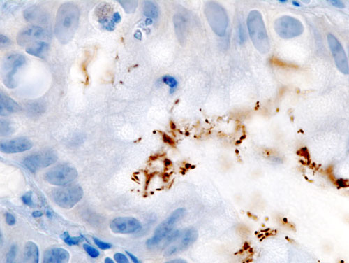
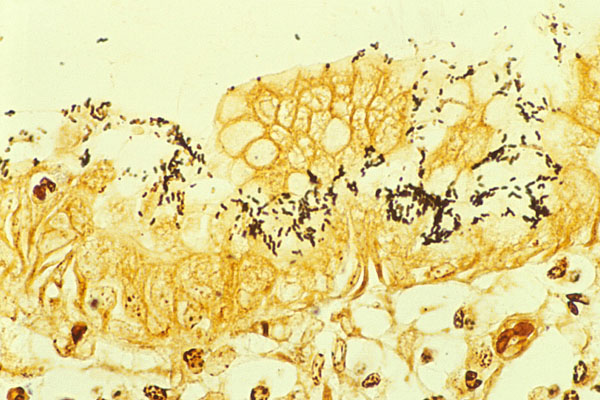
Helicobacter pylori (Template:PronEng) is a gram-negative, microaerophilic, and acidophilic bacterium that infects various areas of the stomach and duodenum. Many cases of peptic ulcers, gastritis, duodenitis, and perhaps some cancers are caused by H. pylori infections. However, many who are infected do not show any symptoms of disease. Helicobacter spp. are the only known microorganisms that can thrive in the highly acidic environment of the stomach. H. pylori's helical shape (from which the genus name is derived) is thought to have evolved to penetrate and favor its motility in the mucus gel layer.[1]
History[edit | edit source]
In 1875, German scientists found helical shaped bacteria in the lining of the human stomach. The bacteria could not be grown in culture and the results were eventually forgotten.[2]
In 1893, the Italian researcher Giulio Bizzozero described helical shaped bacteria living in the acidic environment of the stomach of dogs.[3]
Professor Walery Jaworski of the Jagiellonian University in Kraków investigated sediments of gastric washings obtained from humans in 1899. Among some rod-like bacteria, he also found bacteria with a characteristic helical shape, which he called Vibrio rugula. He was the first to suggest a possible role of this organism in the pathogeny of gastric diseases. This work was included in the "Handbook of Gastric Diseases" but it did not have much impact as it was written in Polish.[4]
The bacterium was rediscovered in 1979 by Australian pathologist Robin Warren, who did further research on it with Barry Marshall beginning in 1981; they isolated the organisms from mucosal specimens from human stomachs and were the first to successfully culture them.[5] In their original paper,[6] Warren and Marshall contended that most stomach ulcers and gastritis were caused by infection by this bacterium and not by stress or spicy food as had been assumed before.[7]
The medical community was slow to recognize the role of this bacterium in stomach ulcers and gastritis, believing that no microorganism could survive for long in the acidic environment of the stomach. The community began to come around after further studies were done, including one in which Marshall drank a Petri dish of H. pylori, developed gastritis, and the bacteria were recovered from his stomach lining, thereby satisfying three out of the four Koch's postulates. The fourth was satisfied after a second endoscopy ten days after inoculation revealed signs of gastritis and the presence of "H. pylori". Marshall was then able to treat himself using a fourteen day dual therapy with bismuth salts and metronidazole. Marshall and Warren went on to show that antibiotics are effective in the treatment of many cases of gastritis. In 1994, the National Institutes of Health (USA) published an opinion stating that most recurrent gastric ulcers were caused by H. pylori, and recommended that antibiotics be included in the treatment regimen.[8] Evidence has been accumulating to suggest that duodenal ulcers are also associated with H. pylori infection.[9][10] In 2005, Warren and Marshall were awarded the Nobel Prize in Medicine for their work on H. pylori.[11]
Before the appreciation of the bacterium's role, stomach ulcers were typically treated with medicines that neutralize gastric acid or decrease its production. While this worked well, the ulcers very often reappeared. A very often used medication against gastritis and peptic ulcers was bismuth subsalicylate. It was often effective, but fell out of use, since its mechanism of action was a mystery. Nowadays it is quite clear that it is due to the bismuth salt acting as an antibiotic. Today, many stomach ulcers are treated with antibiotics effective against H. pylori.
The bacterium was initially named Campylobacter pyloridis, then C. pylori (after a correction to the Latin grammar) and in 1989, after DNA sequencing and other data showed that the bacterium did not belong in the Campylobacter genus, it was placed in its own genus, Helicobacter. The name pylōri means "of the pylorus" or pyloric valve (the circular opening leading from the stomach into the duodenum), from the Greek word πυλωρός, which means gatekeeper.
While H. pylori remains the most medically important bacterial inhabitant of the human stomach, other species of the Helicobacter genus have been identified in other mammals and some birds, and some of these can infect humans.[12] Helicobacter species have also been found to infect the livers of certain mammals and to cause liver disease.[13]
Structure[edit | edit source]
H. pylori is a helical shaped Gram-negative bacterium, about 3 micrometres long with a diameter of about 0.5 micrometre. It has 4–6 flagella. It is microaerophilic, i.e. it requires oxygen but at lower levels than those contained in the atmosphere. It contains a hydrogenase which can be used to obtain energy by oxidizing molecular hydrogen (H2) that is produced by other intestinal bacteria.[14] It tests positive for oxidase, catalase, and urease. It is capable of forming biofilms[15] and conversion from helical to coccoid form[16], both likely to favor its survival and be factors in the epidemiology of the bacterium. The coccoid form of the organism has not been cultured, but has been found in the water supply in the US. This form has also been found to be able to adhere to gastric epithelial cells in vitro.

Colonization[edit | edit source]
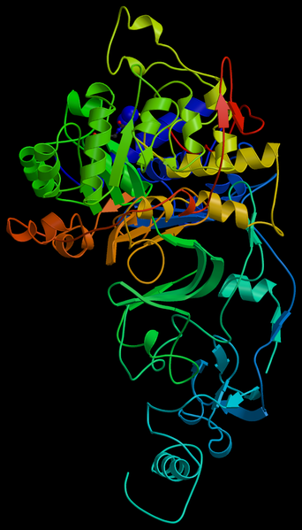
With its flagella, the bacterium moves through the stomach lumen and drills into the mucus gel layer of the stomach. It then finds ways to live in various areas of the stomach. The known areas include: inside the mucus gel layer (with a preference for the superficial area), above epithelial cells, and inside vacuoles formed by H. pylori in epithelial cells. It produces adhesins which bind to membrane-associated lipids and carbohydrates and help its adhesion to epithelial cells. An example of this is the Lewis b antigen. It produces large amounts of urease enzymes which are localized inside and outside of the bacterium. Urease metabolizes urea (which is normally secreted into the stomach) to carbon dioxide and ammonia (which neutralizes gastric acid). The survival of H. pylori in the acidic stomach is dependent on urease, and it would eventually die without it. The ammonia that is produced is toxic to the epithelial cells, and, along with the other products of H. pylori—including protease, catalase and certain phospholipases—causes damage to those cells.
Some strains of the bacterium have a particular mechanism for "injecting" the inflammatory inducing agents peptidoglycan from their own cell wall into epithelial stomach cells. (See below for "cagA pathogenicity island" in the section Genome studies of different strains) This factor may play a role in allowing certain strains to invade host tissue.[17]
Causes of infection[edit | edit source]
H. pylori is a contagious bacterium. Many researchers think that H. pylori is transmitted orally by means of fecal matter through the ingestion of waste tainted food or water. A clean and hygienic environment can help decrease the risk of H. pylori infection.
Diagnosis of infection[edit | edit source]

Diagnosis of infection is usually made by checking for dyspeptic symptoms and then doing tests which can suggest H. pylori infection. One can test noninvasively for H. pylori infection with a blood antibody test, stool antigen test, or with the carbon urea breath test (in which the patient drinks 14C- or 13C-labelled urea, which the bacterium metabolizes producing labelled carbon dioxide that can be detected in the breath). However, the most reliable method for detecting H. pylori infection is a biopsy check during endoscopy with a rapid urease test, histological examination, and microbial culture. None of the test methods is completely failsafe. Even biopsy is dependent on the location of the biopsy. Blood antibody tests, for example, range from 76% to 84% sensitivity. Some drugs can affect H. pylori urease activity and give "false negatives" with the urea-based tests.
Infection may be symptomatic or asymptomatic (without perceptible ill effects). It is estimated that up to 70% of infection is asymptomatic and that about 2/3 of the world population are infected by the bacterium, making it the most widespread infection in the world. Actual infection rates vary from nation to nation - the West (Western Europe, North America, Australasia) having rates around 25% and much higher in the Third World. In the latter, it is common, probably due to poor sanitary conditions, to find infections in children. In the United States, infection is primarily in the older generations (about 50% for those over the age of 60 compared with 20% under 40 years) and the poorest.
This is largely attributed to higher hygiene standards and widespread use of antibiotics. However, antibiotic resistance is appearing in H. pylori.[18] There are already many metronidazole resistant strains in Europe, the United States, and developing countries.
The bacteria have been isolated from feces, saliva and dental plaque of infected patients, which suggests gastro-oral or fecal-oral as possible transmission routes.
It is widely believed that in the absence of treatment, H. pylori infection—once established in its gastric niche—persists for life. In the elderly, however, it is likely infection can disappear as the stomach's mucosa becomes increasingly atrophic and inhospitable to colonization. The proportion of acute infections that persist is not known, but several studies that followed the natural history in populations have reported apparent spontaneous elimination.[19][20]==Gallery==
-
Scanning electron micrograph depicts a Flexispira rappini bacteria (13,951x mag). From Public Health Image Library (PHIL). [21]
-
Scanning electron micrograph depicts a Flexispira rappini bacteria (6976x mag). From Public Health Image Library (PHIL). [21]
-
Scanning electron micrograph depicts a Flexispira rappini bacteria (13951x mag). From Public Health Image Library (PHIL). [21]
Helicobacter and cancer[edit | edit source]
While the incidence of H. pylori infection in humans is decreasing in developing countries, presumably because of improving sanitation and increasing use of antibiotics, in the United States the incidence of gastric cancer has decreased by 80 percent from 1900 to 2000. This apparent correlation is consistent with an epidemiological link between H. pylori and cancer. Specifically, both gastric cancer and gastric MALT lymphoma (lymphoma of the mucosa-associated lymphoid tissue) have been associated with H. pylori, and the bacterium has been categorized as a group I carcinogen by the International Agency for Research on Cancer (IARC). Despite these associations, a direct causal relationship has not been demonstrated. Nonetheless, among bacteria suspected to cause cancer, H. pylori is the leading contender.
Two related mechanisms by which H. pylori could promote cancer are under investigation. One mechanism involves the enhanced production of free radicals near H. pylori and an increased rate of host cell mutation. The other proposed mechanism has been called a "perigenetic pathway"[22] and involves enhancement of the transformed host cell phenotype by means of alterations in cell proteins such as adhesion proteins. It has been proposed that H. pylori induces inflammation and locally high levels of TNF-alpha and/or interleukin 6. According to the proposed perigenetic mechanism, inflammation-associated signaling molecules such as TNF-alpha can alter gastric epithelial cell adhesion and lead to the dispersion and migration of mutated epithelial cells without the need for additional mutations in tumor suppressor genes such as genes that code for cell adhesion proteins.
Acid reflux and esophageal cancer[edit | edit source]
As the incidence of gastric cancer has decreased, the incidences of gastroesophageal reflux disease and esophageal cancer have increased dramatically. In 1996, Martin J. Blaser put forward the theory that H. pylori might also have a beneficial effect: by regulating the acidity of the stomach contents, it lowers the impact of regurgitation of gastric acid into the esophagus.[2] While some favorable evidence has been accumulated, as of 2005 the theory is not universally accepted.
Genome studies of different strains[edit | edit source]

Several strains are known, and the genomes of two have been completely sequenced.[23][24] The genome of the strain "26695" consists of about 1.7 million base pairs, with some 1550 genes. The two sequenced strains show large genetic differences, with up to 6% of the nucleosides differing.
Study of the H. pylori genome is centered on attempts to understand pathogenesis, the ability of this organism to cause disease. There are 62 genes in the "pathogenesis" category of the genome database. Both sequenced strains have an approximately 40 kb long Cag pathogenicity island (a common gene sequence believed responsible for pathogenesis) that contains over 40 genes. This pathogenicity island is usually absent from H. pylori strains isolated from humans who are carriers of H. pylori but remain asymptomatic.
The cagA gene codes for one of the major H. pylori virulence proteins. Bacterial strains that have the cagA gene are associated with an ability to cause severe ulcers. The cagA gene codes for a relatively long (1186 amino acid) protein. The cagA protein is transported into human cells where it may disrupt the normal functioning of the cytoskeleton. The Cag pathogenicity island has about 30 genes that code for a complex type IV secretion system. After attachment of H.pylori to stomach epithelial cells, the cagA protein is injected into the epithelial cells by the type IV secretion system. The cagA protein is phosphorylated on tyrosine residues by a host cell membrane-associated tyrosine kinase. Pathogenic strains of H. pylori have been shown to activate the epidermal growth factor receptor (EGFR), a membrane protein with a tyrosine kinase domain. Activation of the EGFR by H. pylori is associated with altered signal transduction and gene expression in host epithelial cells that may contribute to pathogenesis. It has also been suggested that a c-terminal region of the cagA protein (amino acids 873–1002) can regulate host cell gene transcription independent of protein tyrosine phosphorylation. It is thought, due to cagA's low GC content relative to the rest of the helicobacter genome, that the gene was acquired by horizontal transfer from another cagA+ bacterial species.
Treatment of infection[edit | edit source]

In peptic ulcer patients where infection is detected, the normal procedure is eradicating H. pylori to allow the ulcer to heal. The standard first-line therapy is a one week triple therapy. The Sydney gastroenterolgist Thomas Borody invented the first triple therapy in 1987.[25] Today, the standard triple therapy is amoxicillin, clarithromycin and a proton pump inhibitor such as omeprazole.[26] Variations of the triple therapy have been developed over the years, such as using a different proton pump inhibitor, as with pantoprazole or rabeprazole, or using metronidazole instead of amoxicillin in those allergic to penicillin.[27] Such a therapy has revolutionised the treatment of peptic ulcers and has made a cure to the disease possible, where previously symptom control using antacids, H2-antagonists or proton pump inhibitors alone was the only option.[28][29]
A meta-analysis of randomized controlled trials suggests that supplementation with probiotics can improve eradication rates and reduce adverse events.[30]
Unfortunately, an increasing number of infected individuals are found to harbour antibiotic-resistant bacteria. This results in initial treatment failure and requires additional rounds of antibiotic therapy or alternative strategies such as a quadruple therapy. Bismuth compounds are also effective in combination with the above drugs. For the treatment of clarithromycin-resistant strains of H. pylori the use of levofloxacin as part of the therapy has been suggested.
Some studies show that consumption of broccoli sprouts can be effective at inhibiting H. pylori growth[31] with sulforaphane being at least one of the active agents[32].
Some studies show that mastic gum can destroy H. pylori in vitro, but studies done in vivo have shown it to be ineffective.[33]
A study done on Mongolian gerbils indicates that green tea extract can suppress H. pylori growth.[34] Another study done in South Korea suggests that an acidic polysaccharide found in green tea is significantly effective in preventing adhesion of H. pylori to human cultures of epithelial cells.[35]
As explained below, some authors suggest that some strains of H. pylori may be protective against certain diseases of the esophagus and cardia. Therefore, a more cautious approach than complete eradication may be necessary in some cases.
Treatment Regimen[edit | edit source]
- 1. Peptic ulcer disease[36]
- In patients aged 55 years or younger with no alarm features, two management options may be considered:
- 1.1 Indications for eradication therapy
- In moderate to high prevalence of H. pylori infection (≥ 10%): Test-and-treat strategy using a validated noninvasive test (urea breathing test or stool antigen test)
- In low prevalence situations: Treatment indicated after the empiric trial of acid suppression with a proton pump inhibitor for 4–8 weeks
- 1.2 Proton pump inhibitors (PPI)
- Preferred regimen (1): Lansoprazole 30 mg q12h
- Preferred regimen (2): Omeprazole 20 mg q12h
- Preferred regimen (3): Esomeprazole 40 mg q24h
- Preferred regimen (4): Rabeprazole 20 mg q12h
- 1.3 Regimens for Initial Treatment
- 1.3.1 Triple therapy
- Preferred regimen (1): Proton pump inhibitor standard dose bid AND Amoxicillin 1 g bid AND Clarithromycin 500 mg bid for 7-14 days
- Preferred regimen (2): Proton pump inhibitor standard dose bid AND Amoxicillin 1 g bid AND Metronidazole 500 mg bid for 7-14 days
- Preferred regimen (3) (Levofloxacin triple therapy): Proton pump inhibitor standard dose bid AND Amoxicillin 1 g bid AND Levofloxacin 500 mg bid for 10 days
- Preferred regimen (4) (Rifabutin triple therapy): Proton pump inhibitor standard dose bid AND Amoxicillin 1 g bid AND Rifabutin 150-300 mg/day for 10 days
- 1.3.2 Quadruple therapy
- Preferred regimen (1): Proton pump inhibitor standard dose bid AND Metronidazole 250 mg q6h AND Tetracycline 500 mg q6h AND Bismuth (dose depends on preparation) for 10-14 days
- Preferred regimen (2) non-bismuth quadruple therapy (concomitant therapy): Proton pump inhibitor (standard dose twice daily) for 7–14 days AND Clarithromycin 500 mg bid for 7–14 days AND Amoxicillin 1 g bid for 10 days AND Metronidazole 250 mg qid for 7–14 days
- 1.3.3 Sequential therapy
- Preferred regimen: Proton pump inhibitor standard dose bid AND Amoxicillin 1 g bid for 1-5 days followed by Proton pump inhibitor standard dose bid AND Clarithromycin 500 mg bid AND Tinidazole 500 mg bid for 6-10 days
- Note: Alternative triple therapies appropriate for patients with an allergy to Amoxicillin include (Proton pump inhibitor and Clarithromycin and Metronidazole) or (Proton pump inhibitor and Tetracycline and Metronidazole)
- 1.5 Clarithromycin resistance
- 1.5.1 Clarithromycin resistance ≥ 20%
- Preferred regimen (1) (bismuth quadruple therapy): Proton pump inhibitor standard dose bid AND Metronidazole 250 mg q6h AND Tetracycline 500 mg q6h AND Bismuth (dose depends on preparation) for 10-14 days
- Preferred regimen (2) (sequential therapy): Proton pump inhibitor standard dose bid AND Amoxicillin 1 g bid for 1-5 days followed by Proton pump inhibitor standard dose bid AND Clarithromycin 500 mg bid AND Tinidazole 500 mg bid for 6-10 days
- Preferred regimen (3) non-bismuth quadruple therapy (concomitant therapy): Proton pump inhibitor (standard dose twice daily) for 7–14 days AND Clarithromycin 500 mg bid for 7–14 days AND Amoxicillin 1 g bid for 10 days AND Metronidazole 250 mg qid for 7–14 days
- Alternative regimen: Proton pump inhibitor standard dose bid AND Amoxicillin 1 g bid AND Levofloxacin 500 mg bid for 10 days
- 1.5.2 Clarithromycin resistance < 20%
- Preferred regimen (1): Proton pump inhibitor standard dose bid for 7-14 days AND Clarithromycin 500 mg bid for 7–14 days AND Amoxicillin 1 g bid for 7–14 days OR Metronidazole 250 mg qid for 7–14 days
- Preferred regimen (2): Proton pump inhibitor standard dose bid AND Metronidazole 250 mg q6h AND Tetracycline 500 mg q6h AND Bismuth (dose depends on preparation) for 10-14 days
- Alternative regimen (1):Proton pump inhibitor standard dose bid AND Metronidazole 250 mg q6h AND Tetracycline 500 mg q6h AND Bismuth (dose depends on preparation) for 10-14 days
- Alternative regimen (2): Proton pump inhibitor standard dose bid for 10 days AND Levofloxacin 500 mg bid for 10 days AND Amoxicillin 1 g bid for 10 days
See also[edit | edit source]
Notes[edit | edit source]
- ↑ Chapter 23: Campylobacter and Helicobacter from Medical Microbiology, 4th ed., by Samuel Baron (editor), 1996
- ↑ 2.0 2.1 Blaser MJ (2005). "An Endangered Species in the Stomach". Scientific American. 292 (2): 38–45. PMID 15715390.
- ↑ Bizzozero, Giulio (1893). "Ueber die schlauchförmigen Drüsen des Magendarmkanals und die Beziehungen ihres Epitheles zu dem Oberflächenepithel der Schleimhaut". Archiv für mikroskopische Anatomie. 42: 82–152.
- ↑ Konturek JW (2003 Dec). "Discovery by Jaworski of Helicobacter pylori and its pathogenetic role in peptic ulcer, gastritis and gastric cancer". J Physiol Pharmacol. 54 Suppl 3: 23–41. PMID 15075463. Check date values in:
|year=(help) - ↑ Barry Marshall biography, The Tall Poppy Campaign. Accessed 9 September 2006.
- ↑ Marshall BJ (1983). "Unidentified curved bacillus on gastric epithelium in active chronic gastritis". Lancet. 1 (8336): 1273–1275. PMID 6134060.
- ↑ Marshall BJ, Warren JR (1984). "Unidentified curved bacilli in the stomach patients with gastritis and peptic ulceration". Lancet. 1 (8390): 1311–1315. PMID 6145023.
- ↑ "Helicobacter pylori in peptic ulcer disease". NIH Consensus Statement Online Jan 7–9;12(1):1-23. Retrieved 2004-12-21.
- ↑ Pietroiusti A, Luzzi I, Gomez MJ, Magrini A, Bergamaschi A, Forlini A, Galante A. (2005). "Helicobacter pylori duodenal colonization is a strong risk factor for the development of duodenal ulcer". PMID 15801926. Unknown parameter
|month=ignored (help) - ↑ Helicobacter pylori infection induces duodenitis and superficial duodenal ulcer in Mongolian gerbils
- ↑ The Nobel Prize in Physiology or Medicine 2005 awarded to Barry J. Marshall and J. Robin Warren "for their discovery of the bacterium Helicobacter pylori and its role in gastritis and peptic ulcer disease".
- ↑ Mobley, Harry L. T. (2001). Helicobacter Pylori: Physiology and Genetics. ASM Press. ISBN 1555812139. Unknown parameter
|coauthors=ignored (help) - ↑ Starzyñska T, Malfertheiner P (2006). "Helicobacter and digestive malignancies". Helicobacter. 11 Suppl 1: 32–5. PMID 16925609.
- ↑ Jonathan W. Olson and Robert J. Maier. Molecular Hydrogen as an energy source for Helicobacter pylori. Science 298.5599 (Nov 29, 2002): pp.1788–1790. PMID 12459589
- ↑ Stark RM, Gerwig GJ, Pitman RS, Potts LF, Williams NA, Greenman J, Weinzweig IP, Hirst TR, Millar MR (1999). "Biofilm formation by Helicobacter pylori". PMID 10063642.
- ↑ Chan WY, Hui PK, Leung KM, Chow J, Kwok F, Ng CS (1994). "Coccoid forms of Helicobacter pylori in the human stomach". PMID 7524304.
- ↑ Viala, Jérôme; Chaput, Catherine; Boneca, Ivo G; Cardona, Ana; Girardin, Stephen E; Moran, Anthony P; Athman, Rafika; Memet, Sylvie; Huerre, Michel R; Coyle, Anthony J; DiStefano, Peter S; Sansonetti, Philippe J; Labigne, Agnès; Bertin, John; Philpott, Dana J; Ferrero, Richard L (2004). "Nod1 responds to peptidoglycan delivered by the Helicobacter pylori cag pathogenicity island". Nature Immunol. 5: 1166–74. doi:10.1038/ni1131, PMID 15489856.
- ↑ H pylori antibiotic resistance: prevalence, importance, and advances in testing
- ↑ Goodman K, O'rourke K, Day R, Wang C, Nurgalieva Z, Phillips C, Aragaki C, Campos A, de la Rosa J (2005). "Dynamics of Helicobacter pylori infection in a US-Mexico cohort during the first two years of life". Int J Epidemiol. 34 (6): 1348–55. PMID 16076858.
- ↑ Goodman K, Cockburn M (2001). "The role of epidemiology in understanding the health effects of Helicobacter pylori". Epidemiology. 12 (2): 266–71. PMID 11246592.
- ↑ 21.0 21.1 21.2 "Public Health Image Library (PHIL)".
- ↑ Tsuji S, Kawai N, Tsujii M, Kawano S, Hori M (2003). "Review article: inflammation-related promotion of gastrointestinal carcinogenesis - a perigenetic pathway". Aliment Pharmacol Ther. 18 (Suppl 1): 82–9. PMID 12925144.
- ↑ Tomb JF, White O, Kerlavage AR, Clayton RA, Sutton GG, Fleischmann RD, Ketchum KA, Klenk HP, Gill S, Dougherty BA, Nelson K, Quackenbush J, Zhou LX, Kirkness EF, Peterson S, Loftus B, Richardson D, Dodson R, Khalak HG, Glodek A, McKenney K, Fitzegerald LM, Lee N, Adams MD, Hickey EK, Berg DE, Gocayne JD, Utterback TR, Peterson JD, Kelley JM, Cotton MD, Weldman JM, Fujii C, Bowman C, Watthey L, Wallin E, Hayes WS, Weidman JM, Fujii C, Borodovsky M, Karp PD, Smith HO, Fraser CM, Venter JC (1997). "The complete genome sequence of the gastric pathogen Helicobacter pylori". Nature. 288 (6642): 539–547. PMID 9252185.
- ↑ The Pylori Gene website allows access to genome information for the H. pylori 26695 and J99 strains. Another genome interface is provided by the National Center for Biotechnology Information; 26695, J99.
- ↑ Borody, Thomas J. (October 16, 1989). "Recurrence of duodenal ulcer and Campylobacter pylori infection after eradication". Medical Journal of Australia. 151 (8): 431–435. PMID 2687668. Text " P. Cole, S. Noonan, A. Morgan, J. Lenne, L. Hyland, S. Brandl, E. G. Borody, and L. L. George " ignored (help); Check date values in:
|date=(help) - ↑ Mirbagheri, Seyed Amir (August 14, 2006). "Triple, standard quadruple and [[ampicillin]]-[[sulbactam]]-based quadruple therapies for H pylori eradication: A comparative three-armed randomized clinical trial". World Journal of Gastroenterology. 12 (30): 4888–4891. PMID 16937475. Retrieved 2006-09-30. Unknown parameter
|coauthors=ignored (help); Check date values in:|date=(help); URL–wikilink conflict (help) - ↑ European Helicobacter Pylori Study Group. "Current Concepts in the Management of Helicobacter pylori Infection". The Maastricht 2-2000 Consensus Report. Unknown parameter
|accessyear=ignored (|access-date=suggested) (help); Unknown parameter|accessmonthday=ignored (help) - ↑ Rauws EA, Tytgat GN (1990). "Cure of duodenal ulcer associated with eradication of Helicobacter pylori". Lancet. 335 (8700): 1233–5. PMID 1971318.
- ↑ Graham DY, Lew GM, Evans DG, Evans DJ, Klein PD (1991). "Effect of triple therapy (antibiotics plus bismuth) on duodenal ulcer healing. A randomized controlled trial". Ann. Intern. Med. 115 (4): 266–9. PMID 1854110.
- ↑ Tong JL, Ran ZH, Shen J, Zhang CX, Xiao SD (2007). "Meta-analysis: the effect of supplementation with probiotics on eradication rates and adverse events during Helicobacter pylori eradication therapy". Aliment. Pharmacol. Ther. 25 (2): 155–68. doi:10.1111/j.1365-2036.2006.03179.x. PMID 17229240. ACP Journal Club
- ↑ Galan MV, Kishan AA, Silverman AL (2004). "Oral broccoli sprouts for the treatment of Helicobacter pylori infection: a preliminary report". Dig Dis Sci. 49 (7–8): 1088–90. PMID 15387326. Unknown parameter
|month=ignored (help) - ↑ Fahey JW, Haristoy X, Dolan PM, Kensler TW, Scholtus I, Stephenson KK, Talalay P, Lozniewski A (2002). "Sulforaphane inhibits extracellular, intracellular, and antibiotic-resistant strains of Helicobacter pylori and prevents benzo[a]pyrene-induced stomach tumors". PMID 12032331. Unknown parameter
|month=ignored (help) - ↑ PubMed search for "gum mastic" and "H. pylori", accessed 9 September 2006.
- ↑ Matsubara S, Shibata H, Ishikawa F, Yokokura T, Takahashi M, Sugimura T, Wakabayashi K. (2003). "Suppression of Helicobacter pylori-induced gastritis by green tea extract in Mongolian gerbils". PMID 14550260. Unknown parameter
|month=ignored (help) - ↑ Lee J, Shim J, Lee J, Kim J, Yang I, Chung M, Kim K (2006). "Inhibition of pathogenic bacterial adhesion by acidic polysaccharide from green tea (Camellia sinensis)". J Agric Food Chem. 54 (23): 8717–23. PMID 17090112.
- ↑ Lua error: expandTemplate: template "citation error" does not exist.
References[edit | edit source]
- "Helicobacter pylori and peptic ulcer". Centers for Disease Control and Prevention. Retrieved 2004-12-21.
- Logan, Robert P H; Walker, Marjorie M (20 October 2001). "Clinical review: ABC of the upper gastrointestinal tract. Epidemiology and diagnosis of Helicobacter pylori infection". BMJ. 323: 920–922. PMID 11668141.
External links[edit | edit source]
| Wikimedia Commons has media related to Helicobacter pylori. |
- Helicobacter pylori - Natural solutions Offers guidance and natural programmes for H Pylori sufferers
- The Helicobacter Foundation provides information on Helicobacter pylori and its effects - founded by Dr. Barry J. Marshall, one of the discoverers of H. pylori
- Helicobacter pylori persistence: biology and disease by Martin J. Blaser and John C. Atherton in Journal of Clinical Investigations (2004) Volume 113 pages 321–333.
- Helicobacter pylori: Physiology and Genetics Online text of the Mobley et al book (via NCBI).
- European Helicobacter Study Group sponsors annual international workshop, yearbook, and clinical guidelines
- The Year in Helicobacter pylori 2006 Helicobacter journal Supplement Oct 2006 - Free content
- 1994–2005 back issues
- Atwood KC. Bacteria, Ulcers, and Ostracism?: H. pylori and the Making of a Myth. Skeptical Inquirer November 2004. Fulltext Claims that medicine’s purported ostracism of the discovery of H. pylori has achieved a mythological quality, but isn't true: after appropriate initial scientific skepticism, the hypothesis was accepted right "on schedule".
- Microscopic image of H. pylori
- H. pylori Picture Book
- List of species in the genus Helicobacter
- Out Of Africa -- Bacteria, As Well: Homo Sapiens And H. Pylori Jointly Spread Across The Globe ScienceDaily (Feb. 16, 2007) — When man made his way out of Africa some 60,000 years ago to populate the world, he was not alone: He was accompanied by the bacterium Helicobacter pylori
zh-min-nan:Helicobacter pylori be-x-old:Helicobacter pylori bg:Helicobacter pylori ca:Helicobacter pylori da:Helicobacter pylori de:Helicobacter pylori ko:헬리코박터 파일로리 it:Helicobacter pylori he:Helicobacter pylori la:Helicobacter Pylori lb:Helicobacter pylori nl:Helicobacter pylori new:हेलिकोब्याक्टर पाइलोरी no:Helicobacter pylori nn:Helicobacter pylori sk:Helicobacter pylori sr:Хеликобактер пилори fi:Helicobacter pylori sv:Helicobacter pylori uk:Helicobacter pylori
 KSF
KSF![Scanning electron micrograph depicts a Flexispira rappini bacteria (13,951x mag). From Public Health Image Library (PHIL). [21]](https://www.wikidoc.org/images/5/50/Helicobacter_pylori05.jpeg)
![Scanning electron micrograph depicts a Flexispira rappini bacteria (6976x mag). From Public Health Image Library (PHIL). [21]](https://www.wikidoc.org/images/1/11/Helicobacter_pylori04.jpeg)
![Scanning electron micrograph depicts a Flexispira rappini bacteria (13951x mag). From Public Health Image Library (PHIL). [21]](https://www.wikidoc.org/images/4/41/Helicobacter_pylori03.jpeg)