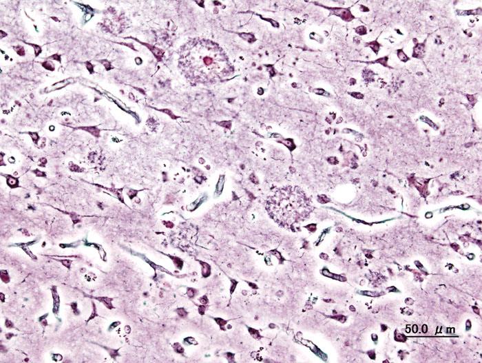
Senile plaques
 From Wikidoc - Reading time: 3 min
|
WikiDoc Resources for Senile plaques |
|
Articles |
|---|
|
Most recent articles on Senile plaques Most cited articles on Senile plaques |
|
Media |
|
Powerpoint slides on Senile plaques |
|
Evidence Based Medicine |
|
Clinical Trials |
|
Ongoing Trials on Senile plaques at Clinical Trials.gov Trial results on Senile plaques Clinical Trials on Senile plaques at Google
|
|
Guidelines / Policies / Govt |
|
US National Guidelines Clearinghouse on Senile plaques NICE Guidance on Senile plaques
|
|
Books |
|
News |
|
Commentary |
|
Definitions |
|
Patient Resources / Community |
|
Patient resources on Senile plaques Discussion groups on Senile plaques Patient Handouts on Senile plaques Directions to Hospitals Treating Senile plaques Risk calculators and risk factors for Senile plaques
|
|
Healthcare Provider Resources |
|
Causes & Risk Factors for Senile plaques |
|
Continuing Medical Education (CME) |
|
International |
|
|
|
Business |
|
Experimental / Informatics |
Senile plaques (syn. neuritic plaques, senile druse, braindruse) are extracellular deposits of amyloid in the gray matter of the brain. The deposits are associated with degenerative neural structures and an abundance of microglia and astrocytes. Large numbers of senile plaques and neurofibrillary tangles are characteristic features of Alzheimer’s disease.

The plaques are variable in shape and size, but are on the average 50 µm in size. (Franke, M.) In Alzheimer's disease they are primarily composed of amyloid beta peptides. These polypetides tend to aggregate and are believed to be neurotoxic.
Verification[edit | edit source]
Senile plaques are visible in light microscopy after staining by silver, Congo red, Thioflavin, Kresylviolett, PAS-reaction, and by fluorescence and immunofluorescence microscopy.
Occurrence[edit | edit source]
Senile plaques can be found in human and animal brains (e.g. mammals and birds). From an age of 60 years (10%) to an age of 80 years (60%) the proportion of people with plaques increases approximately linearly. A small number of plaques can be due to the physiological process of aging. Women are slightly more likely to have plaques than males (Franke, M.). The plaques occur commonly in the amygdoid nucleus and the sulci of the cortex of brain.
History[edit | edit source]
Blocq and Marinesco first described plaques in the grey matter in 1892. Because of their similarity to the actinomyces druses they were called druse necrosis by O. Fischer in the beginning of the 20th century. The connection of plaques and demential illness was discovered by Alzheimer in 1906. Bielschowsky supposed in 1911 the amyloid-nature of the plaques. Wisniewski denominated them neuritic plaques in 1973. The second half of the 20th century saw proposed theories of immunological and genetic factors in plaque formation (Katenkamp, Op den Velde und Stam). Statistically investigations were performed by J.A.N. Corsellis and M. Franke in the 1970s. M. Franke showed that a demential disease is likely when the number of senile plaques in the frontal cortex is more than 200/mm3. In 1985 succeeded the biochemical identification of amyloid beta. But there are more unsolved questions of formation and importance of the plaque formation.
Importance[edit | edit source]
The senile plaques are an important criterion of the neuropathological-histological verification of the Alzheimer’s disease; other factors in verification include pathological neurofibrillaries, tangles, atrophic brain with hydrocephalus, and other degenerative signs. The formation and the distribution of the pathological neurofibrillaries have a regularity (H. Braak and E. Braak) and allows to stage the disease. In combination with the occurrence of a great number of plaques the Alzheimer’s disease can diagnosed with high probability.
Literature[edit | edit source]
- FRANKE, MANFRED: Statistische Untersuchungen über die senilen Drusen im menschlichen Gehirn (zum Problem der Hirndrusenkrankheit). Statistical investigation of the senile plaques in human brains - 1975. Berlin, Akad. für Ärztl. Fortbildung d. DDR, Diss. B, 1976. Deutsche Nationalbiografie Signatur: DBF H 76/10016, IDN: 801313783 Thesen zur Dissertation
- BRAAK, H., E. BRAAK, J. BOHL: Staging of Alzheimer-related cortical destruction. European Neurology, Basel, 1993, 33: 403-408.
- JELLINGER, K.A.: Neurodegenerative Erkrankungen (ZNS) - Eine aktuelle Übersicht. Journal für Neurologie, Neurochirurgie und Psychiatrie 2005; 6 (1), 9-18
- CRUZ, L.; B. URBANC: Aggregation and disaggregation of senile plaques in Alzheimer disease. Proc. Natl. Acad. Sci. USA Vol. 94, pp. 7612±7616, July 1997 Neurobiology
 KSF
KSF