Subarachnoid hemorrhage CT
 From Wikidoc - Reading time: 4 min
|
Subarachnoid Hemorrhage Microchapters |
|
Diagnosis |
|---|
|
Treatment |
|
AHA/ASA Guidelines for the Management of Aneurysmal Subarachnoid Hemorrhage (2012)
|
|
Case Studies |
|
Subarachnoid hemorrhage CT On the Web |
|
American Roentgen Ray Society Images of Subarachnoid hemorrhage CT |
|
Risk calculators and risk factors for Subarachnoid hemorrhage CT |
Editor-In-Chief: C. Michael Gibson, M.S., M.D. [1]; Associate Editor(s)-in-Chief: Syed Ahsan Hussain, M.D.[2] Cafer Zorkun, M.D., Ph.D. [3]; Sara Mehrsefat, M.D. [4]
Overview[edit | edit source]
The modality of choice for diagnosis of subarachnoid hemorrhage is noncontrast head computed tomography (CT), with or without lumbar puncture[1] The sensitivity of CT to the presence of subarachnoid blood is strongly influenced by both the amount of blood and the time since the hemorrhage[2]
CT[edit | edit source]
The modality of choice for diagnosis of subarachnoid hemorrhage is noncontrast head computed tomography (CT), with or without lumbar puncture.[1] The diagnosis of subarachnoid hemorrhage cannot be made on clinical grounds alone. Medical imaging is usually required to confirm or exclude bleeding.[3][4]
- The sensitivity of CT to the presence of subarachnoid blood is strongly influenced by both the amount of blood and the time since the hemorrhage[2]
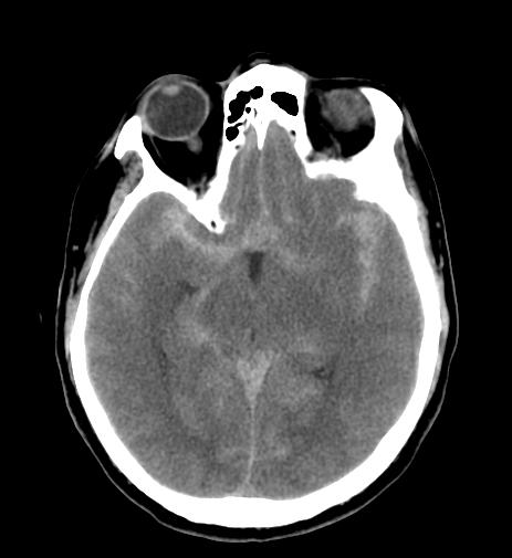
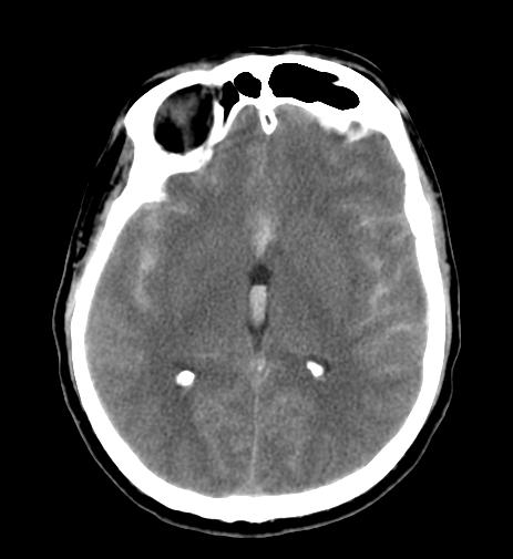
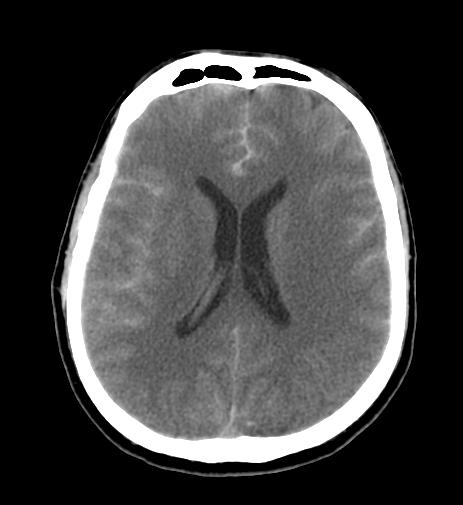
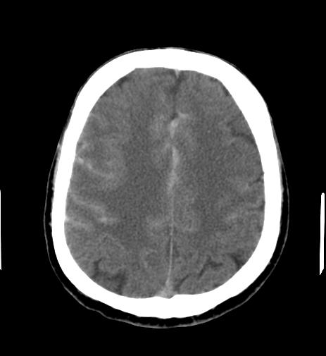
The diagnosis is suspected when hyperattenuating material is seen filling the subarachnoid space. Most commonly this is apparent around
- Circle of Willis (account of the majority of berry aneurysms)
- Sylvian fissure
Small amounts of blood can sometimes be appreciated pooling in the interpeduncular fossa, appearing as a small hyperdense triangle, or within the occipital horns of the lateral ventricles.[5]
Sensitivity of CT may be reduced in the following conditions:[6]
- Patients with atypical symptoms (isolated neck pain)
- Minor bleeds
Subarachnoid haemorrhages are grouped into four categories according to the amount of blood by the Fisher Grade.[7]
| Grading | Amount of blood shown on initial CT scans |
|---|---|
| Grade 1 |
|
| Grade 2 |
|
| Grade 3 |
|
| Grade 4 |
|
Acute[edit | edit source]
- The sensitivity of CT in the first 3 days after aSAH is very high (close to 100%)[8]
- Acute Subarachnoid hemorrhage is typically 50-60 HU
Several days to weeks[edit | edit source]
- When CT scanning is performed several days to weeks after the initial bleed, the findings are more subtle
- The initial high-attenuation of blood and clot tend to decrease, and these appear as intermediate gray
- These findings can be isointense relative to normal brain parenchyma
Images[edit | edit source]
The following are the CT scans associated with diffuse subarachnoid hemorrhage.[9]
References[edit | edit source]
- ↑ 1.0 1.1 van der Wee N, Rinkel GJ, Hasan D, van Gijn J (1995). "Detection of subarachnoid haemorrhage on early CT: is lumbar puncture still needed after a negative scan?". J Neurol Neurosurg Psychiatry. 58 (3): 357–9. PMC 1073376. PMID 7897421.
- ↑ 2.0 2.1 Sames TA, Storrow AB, Finkelstein JA, Magoon MR (1996). "Sensitivity of new-generation computed tomography in subarachnoid hemorrhage". Acad Emerg Med. 3 (1): 16–20. PMID 8749962.
- ↑ Mayberg MR, Batjer HH, Dacey R, Diringer M, Haley EC, Heros RC, Sternau LL, Torner J, Adams HP Jr, Feinberg W, Thies W. Guidelines for the management of aneurysmal subarachnoid hemorrhage: a statement for healthcare professionals from a special writing group of the Stroke Council, American Heart Association. Circulation. 1994;90: 2592–2605.
- ↑ Bederson JB, Connolly ES Jr, Batjer HH, Dacey RG, Dion JE, Diringer MN, Duldner JE Jr, Harbaugh RE, Patel AB, Rosenwasser RH. Guidelines for the management of aneurysmal subarachnoid hemor- rhage: a statement for healthcare professionals from a special writing group of the Stroke Council, American Heart Association [published correction appears in Stroke. 2009;40:e518]. Stroke. 2009;40:994 –1025.
- ↑ Brant WE, Helms C. Fundamentals of Diagnostic Radiology. LWW. (2012) ISBN:1608319113
- ↑ Leblanc R (1987). "The minor leak preceding subarachnoid hemorrhage". J Neurosurg. 66 (1): 35–9. doi:10.3171/jns.1987.66.1.0035. PMID 3783257.
- ↑ Fisher C, Kistler J, Davis J (1980). "Relation of cerebral vasospasm to subarachnoid hemorrhage visualized by computerized tomographic scanning". Neurosurgery. 6 (1): 1–9. PMID 7354892.
- ↑ Perry JJ, Stiell IG, Sivilotti ML, Bullard MJ, Emond M, Symington C; et al. (2011). "Sensitivity of computed tomography performed within six hours of onset of headache for diagnosis of subarachnoid haemorrhage: prospective cohort study". BMJ. 343: d4277. doi:10.1136/bmj.d4277. PMC 3138338. PMID 21768192. Review in: Evid Based Med. 2012 Feb;17(1):27-8
- ↑ Rads wiki, Images courtesy of RadsWiki Images courtesy of RadsWiki
 KSF
KSF


