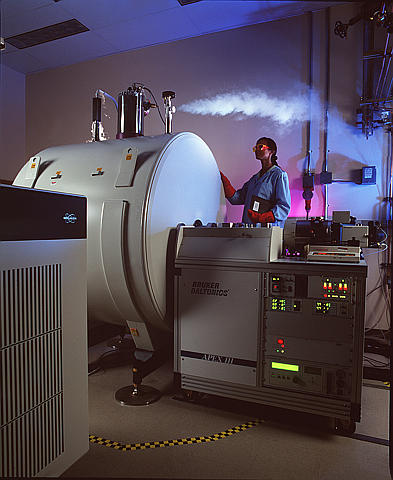
Tandem mass spectrometry
 From Wikidoc - Reading time: 8 min
Tandem mass spectrometry, also known as MS/MS, involves multiple steps of mass spectrometry selection, with some form of fragmentation occurring in between the stages.[1]
Tandem MS instruments[edit | edit source]
Multiple stages of mass analysis separation can be accomplished with individual mass spectrometer elements separated in space or in a single mass spectrometer with the MS steps separated in time. In tandem mass spectrometry in space, the separation elements are physically separated and distinct, although there is a connection between the elements to maintain high vacuum. These elements can be sectors, transmission quadrupole, or time-of-flight. In a tandem mass spectrometry in time instrument, the separation is accomplished with ions trapped in the same place, with multiple separation steps taking place over time. A quadrupole ion trap or FTMS instrument can be used for such an analysis. Trapping instruments can perform multiple steps of analysis, which is sometimes referred to as MSn (MS to the n). Often the number of steps, n, is not indicated, but occasionally the value is specified; for example MS3 indicates three stages of separation.
Notation[edit | edit source]
For tandem mass spectrometry in space, the different elements are often noted in shorthand.
- Q - Quadrupole mass analyzer
- q - Radio frequency collision quadrupole
- TOF - Time-of-flight mass analyzer
- B - Magnetic sector
- E - Electric sector.
The notation can be combined to indicate various hybrid instrument, for example
- QqQ - Triple quadrupole mass spectrometer
- QTOF - Quadrupole time-of-flight mass spectrometer (also QqTOF)
- BEBE - Four-sector (reverse geometry) mass spectrometer.
Tandem MS experiments[edit | edit source]
There are a number of different tandem MS experiments, which each have their own applications and offer their own information. An instrument equipped for tandem MS can still be used to run MS experiments. Tandem MS can be done in either time or space. Tandem MS in space involves the physical separation of the instrument components (QqQ or QTOF), tandem MS in time involves the use of an ion trap.
Tandem MS modes[edit | edit source]
There are four main scan experiments possible using MS/MS.
- Product ion scan
- A precursor ion is selected in the first stage, allowed to fragment and then all resultant masses are detected in the second mass analyzer. This experiment is commonly performed to identify transitions used for quantification by tandem MS.
- Precursor ion scan
- The product ion is selected in the second mass analyzer, and the precursor masses are scanned in the first mass analyzer. A precursor ion scan cannot be done with time based MS instruments.
- Neutral loss scan
- The first mass analyzer scans all the masses. The second mass analyzer also scans, but at a set offset from the first mass analyzer. This offset corresponds to a neutral loss that is commonly observed for the class of compounds. Neutral loss scans cannot be done with time based MS instruments either.
- Selected reaction monitoring
- Both mass analyzers are set to a selected mass. This mode is analogous to selected ion monitoring for MS experiments. A very selective analysis mode, which can increase sensitivity.[2]
Fragmentation in tandem mass spectrometry[edit | edit source]
Fragmentation of gas-phase ions is essential to tandem mass spectrometry and occurs between different stages of mass analysis. There are many methods used to fragment the ions and can result in different types of fragmentation and thus different information about the structure and composition of the molecule.
In-source fragmentation[edit | edit source]
Often, the ionization process is sufficiently violent to leave the resulting ions with sufficient internal energy to fragment within the mass spectrometer. If the product ions persist in their non-equilibrium state for a moderate amount of time before auto-dissociation this process is called metastable fragmentation.[3] [4] Nozzle-skimmer fragmentation refers to the purposeful induction of in-source fragmentation by increasing the nozzle-skimmer potential on usually electrospray based instruments. Although in-source fragmentation allows for fragmentation analysis, it is not technically tandem mass spectrometry unless metastable ions are mass analyzed or selected before auto-dissociation and a second stage of analysis is performed on the resulting fragments. In-source fragmentation is often used in addition to tandem mass spectrometry (with post-source fragmentation) to allow for two steps of fragmentation in a pseudo MS3-type of experiment.[5]
Post-source fragmentation[edit | edit source]

Post-source fragmentation is most often what is being used in a tandem mass spectrometry experiment. Energy can also be added to the, usually already vibrationally excited, ions through post-source collisions with neutral atoms or molecules, the absorption of radiation, or the transfer or capture of an electron by a multiply charged ion. Collision-induced dissociation (CID), also called collisionally activated dissociation (CAD), involves the collision of an ion with a neutral atom or molecule in the gas phase and subsequent dissociation of the ion.[6][7] For example, consider
- <math>AB^+ + M \to A + B^+ + M</math>
where the ion <math>AB^+</math> collides with the neutral species M and subsequently breaks apart. The details of this process are described by collision theory.
If an electron is added to a multiply-charged positive ion, the Coulomb energy is liberated. Adding a free electron is called electron capture dissociation (ECD),[8] and is represented by
- <math>[M + nH]^{n+} + e^- \to \bigg[ [M + (n-1)H]^{(n-1)+} \bigg]^* \to fragments</math>
for a multiply-protonated molecule M. Adding an electron through an ion-ion reaction is called electron transfer dissociation (ETD),[9] and is represented by
- <math>[M + nH]^{n+} + A^- \to \bigg[ [M + (n-1)H]^{(n-1)+} \bigg]^* + A \to fragments</math>.
The energy required for dissociation can be added by photon absorption, resulting in ion photodissociation and represented by
- <math>AB^+ + h\nu \to A + B^+</math>
where <math>h\nu</math> represents the photon absorbed by the ion. Ultraviolet lasers can be used, but can lead to excessive fragmentation of biomolecules.[10] Infrared photons will heat the ions and cause dissociation if enough of them are absorbed. This process is called infrared multiphoton dissociation (IRMPD) and is often accomplished with a carbon dioxide laser and an ion trapping mass spectrometer such as a FTMS.[11] Blackbody radiation can also be used in a technique known as blackbody infrared radiative dissociation (BIRD).[12] In the BIRD method, the entire mass spectrometer vacuum chamber is heated to create infrared radiation.
Peptide fragmentation[edit | edit source]
A peptide sequence tag obtained by tandem mass spectrometry can be used to identify a peptide in a protein database.[14][15][16] A notation has been developed for indicating peptide fragments that arise from a tandem mass spectrum.[13] Peptide fragment ions are indicated by a, b, or c if the charge is retained on the N-terminus and by x, y or z if the charge is maintained on the C-terminus. The subscript indicates the number of amino acid residues in the fragment. Superscripts are sometimes used to indicate neutral losses in addition to the backbone fragmentation, * for loss of ammonia and ° for loss of water. Although peptide backbone cleavage is the most useful for sequencing and peptide identification other fragment ions may be observed under certain conditions. These include the side chain loss ions d, v, w and immonium ions. [17][18]
Oligosaccharide fragmentation[edit | edit source]
Oligosaccharides may be sequenced using tandem mass spectrometry in a similar manner to peptide sequencing. Fragmentation generally occurs on either side of the glycosidic bond (b, c, y and z ions) but also under more energetic conditions through the sugar ring structure in a cross-ring cleavage (x ions). Again trailing subscripts are used to indicate position of the cleavage along the chain. For cross ring cleavage ions the nature of the cross ring cleavage is indicated by preceding superscripts.[19][20]
See also[edit | edit source]
- Chemical kinetics
- Activation energy
- Cross section (physics)
- Shotgun proteomics
- Top-down proteomics
- Newborn screening
References[edit | edit source]
- ↑ IUPAC gold book definition of tandem mass spectrometer [1]
- ↑ deHoffman, Edmond (2003). Mass Spectrometry: Principles and Applications. Toronto: Wiley. p. 133. ISBN 0471485667. Unknown parameter
|coauthors=ignored (help) - ↑ IUPAC gold book definition of metastable ion (in mass spectrometry) [2]
- ↑ IUPAC gold book definition of transient (chemical) species [3]
- ↑ JAMS Vol. 7, Feb. 1996, pp 150-156 [4]
- ↑ Wells JM, McLuckey SA (2005). "Collision-induced dissociation (CID) of peptides and proteins". Meth. Enzymol. 402: 148–85. doi:10.1016/S0076-6879(05)02005-7. PMID 16401509.
- ↑ Sleno L, Volmer DA (2004). "Ion activation methods for tandem mass spectrometry". Journal of mass spectrometry : JMS. 39 (10): 1091–112. doi:10.1002/jms.703. PMID 15481084.
- ↑ Cooper HJ, Håkansson K, Marshall AG (2005). "The role of electron capture dissociation in biomolecular analysis". Mass spectrometry reviews. 24 (2): 201–22. doi:10.1002/mas.20014. PMID 15389856.
- ↑ Syka JE, Coon JJ, Schroeder MJ, Shabanowitz J, Hunt DF (2004). "Peptide and protein sequence analysis by electron transfer dissociation mass spectrometry". Proc. Natl. Acad. Sci. U.S.A. 101 (26): 9528–33. doi:10.1073/pnas.0402700101. PMID 15210983.
- ↑ Morgan JW, Hettick JM, Russell DH (2005). "Peptide sequencing by MALDI 193-nm photodissociation TOF MS". Meth. Enzymol. 402: 186–209. doi:10.1016/S0076-6879(05)02006-9. PMID 16401510.
- ↑ Little DP, Speir JP, Senko MW, O'Connor PB, McLafferty FW (1994). "Infrared multiphoton dissociation of large multiply charged ions for biomolecule sequencing". Anal. Chem. 66 (18): 2809–15. PMID 7526742.
- ↑ Schnier PD, Price WD, Jockusch RA, Williams ER (1996). "Blackbody Infrared Radiative Dissociation of Bradykinin and Its Analogues: Energetics, Dynamics, and Evidence for Salt-Bridge Structures in the Gas Phase". 118 (30): 7178–7189. doi:10.1021/ja9609157. PMID 16525512.
- ↑ 13.0 13.1 Roepstorff P, Fohlman J (1984). "Proposal for a common nomenclature for sequence ions in mass spectra of peptides". Biomed. Mass Spectrom. 11 (11): 601. doi:10.1002/bms.1200111109. PMID 6525415.
- ↑ Hardouin J (2007). "Protein sequence information by matrix-assisted laser desorption/ionization in-source decay mass spectrometry". Mass spectrometry reviews. 26 (5): 672–82. doi:10.1002/mas.20142. PMID 17492750.
- ↑ Shadforth I, Crowther D, Bessant C (2005). "Protein and peptide identification algorithms using MS for use in high-throughput, automated pipelines". Proteomics. 5 (16): 4082–95. doi:10.1002/pmic.200402091. PMID 16196103.
- ↑ Mørtz E, O'Connor PB, Roepstorff P, Kelleher NL, Wood TD, McLafferty FW, Mann M (1996). "Sequence tag identification of intact proteins by matching tanden mass spectral data against sequence data bases". Proc. Natl. Acad. Sci. U.S.A. 93 (16): 8264–7. PMID 8710858.
- ↑ Richard S. Johnson, Stephen A. Martin and Klaus Biemann, Collision-induced fragmentation of (M + H)+ ions of peptides. Side chain specific sequence ions, International Journal of Mass Spectrometry and Ion Processes, Volume 86, 29 December 1988, Pages 137-154. [5]
- ↑ A. M. Falick, W. M. Hines, K. F. Medzihradszky, M. A. Baldwin and B. W. Gibson, Low-mass ions produced from peptides by high-energy collision-induced dissociation in tandem mass spectrometry, Journal of the American Society for Mass Spectrometry, Volume 4, Issue 11, November 1993, Pages 882-893. [6]
- ↑ Bruno Domon, Catherine E Costello (1988). "A systematic nomenclature for carbohydrate fragmentations in FAB-MS/MS spectra of glycoconjugates". Glycoconj. J. 5 (4): 397–409. doi:10.1007/BF01049915.
- ↑ Spina E, Cozzolino R, Ryan E, Garozzo D (2000). "Sequencing of oligosaccharides by collision-induced dissociation matrix-assisted laser desorption/ionization mass spectrometry". Journal of mass spectrometry : JMS. 35 (8): 1042–8. doi:10.1002/1096-9888(200008)35:8<1042::AID-JMS33>3.0.CO;2-Y. PMID 10973004.
Bibliography[edit | edit source]
- McLuckey, Scott A.; Busch, Kenneth L.; Glish, Gary L. (1988). Mass spectrometry/mass spectrometry: techniques and applications of tandem mass spectrometry. New York, N.Y: VCH Publishers. ISBN 0-89573-275-0.
- McLuckey, Scott A.; Glish, Gary L. Mass Spectrometry/Mass Spectrometry: Techniques and Applications of Tandem. Chichester: John Wiley & Sons. ISBN 0-471-18699-6.
- McLafferty, Fred W. (1983). Tandem mass spectrometry. New York: Wiley. ISBN 0-471-86597-4.
- Sherman, Nicholas E.; Kinter, Michael (2000). Protein sequencing and identification using tandem mass spectrometry. New York: John Wiley. ISBN 0-471-32249-0.
 KSF
KSF