Urinary bladder
 From Wikidoc - Reading time: 3 min
Template:Infobox Anatomy Template:Search infobox Steven C. Campbell, M.D., Ph.D.
Overview
[edit | edit source]In anatomy, the urinary bladder is a hollow, muscular, and distensible (or elastic) organ that sits on the pelvic floor in mammals. It is the organ that collects urine excreted by the kidneys prior to disposal by urination. Urine enters the bladder via the ureters and exits via the urethra.
In males, the bladder is superior to the prostate, and separated from the rectum by the rectovesical excavation.
In females, the bladder is separated from the rectum by the rectouterine excavation, and it is separated from the uterus by the vesicouterine excavation.
Regions
[edit | edit source]- Trigone of urinary bladder: The ureters enter the bladder diagonally from its dorsolateral floor in an area called the trigone, which is a triangular shaped area on the postero-inferior wall of the bladder. The urethra exits at the lowest point of the triangle of the trigone.
- Apex: The Median umbilical ligament connects to the apex of the bladder.
- Neck: The Neck is connected to the pubic bone by the pubovesical ligament in women, and by the puboprostatic ligament in men.
Wall
[edit | edit source]The wall of the urinary bladder consists of three layers:
- Mucosa: in this instance transitional epithelium & lamina propria
- Detrusor muscle: consists of an inner and outer longitudinal layer and a middle circular layer of smooth muscle
- a fibrous adventitia and the visceral peritoneum: lie on superior surface
Detrusor muscle
[edit | edit source]The detrusor muscle is a layer of the urinary bladder wall made of smooth muscle fibers arranged in spiral, longitudinal, and circular bundles. When the bladder is stretched, this signals the parasympathetic nervous system to contract the detrusor muscle. This encourages the bladder to expel urine through the urethra.
For the urine to exit the bladder, both the autonomically controlled internal sphincter and the voluntarily controlled external sphincter must be opened. Problems with these muscles can lead to incontinence.
The urinary bladder usually holds 400–620 mL of urine, but it can hold twice this without rupturing if, for example, the outflow is obstructed.
The desire to urinate usually starts when the bladder reaches around 75% of its working volume. If the subject is distracted the desire can fade and return with more urgency as the bladder continues to fill.
See also
[edit | edit source]- Artificial bladder
- Bladder cancer
- Bladder sphincter dyssynergia, a condition in which the sufferer cannot coordinate relaxation of the urethra sphincter with the contraction of the bladder muscles
- Cystitis
- Hematuria, or presence of blood in the urine, is a reason to seek medical attention without delay, as it is a symptom of bladder cancer as well as bladder and kidney stones.
- Neurogenic bladder
- Ureterocele
- Urinary incontinence
- Urodynamics The study of the functional aspects of the detrusor muscle.
- Uvula of urinary bladder
- Vesicouretic reflux
External links
[edit | edit source]- Histology at KUMC epithel-epith09 "Urinary Bladder"
- Template:UCDavisOrganology - "Mammal, bladder (LM, Medium)"
- Template:IowaHistologyInteractive
- Template:SUNYAnatomyLabs - "The Female Pelvis: The Urinary bladder"
- Template:SUNYAnatomyLabs - "The Male Pelvis: The Urinary bladder"
Additional images
[edit | edit source]-
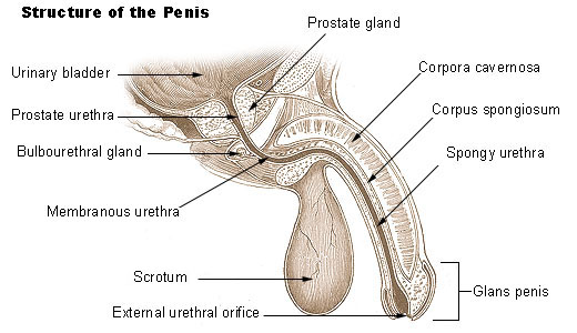 Structure of the penis
 -
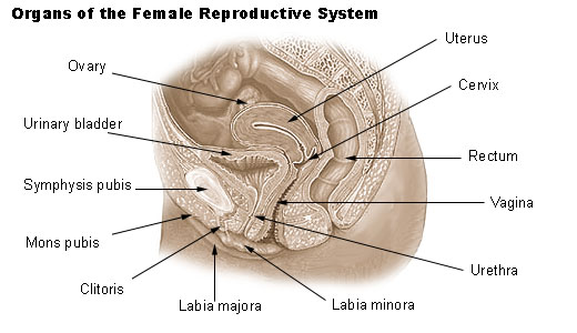 Organs of the female reproductive system.
 -
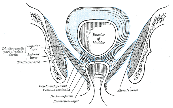 Coronal section of pelvis, showing arrangement of fasciæ. Viewed from behind.
 -
-
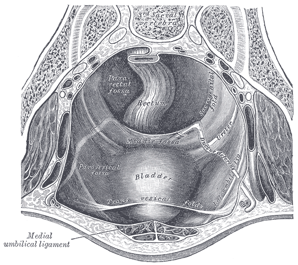 The peritoneum of the male pelvis.
 -
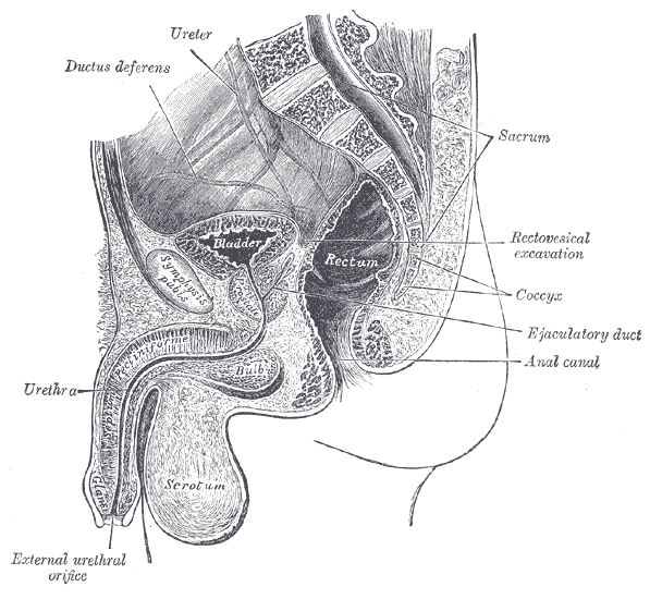 Median sagitta section of male pelvis.
 -
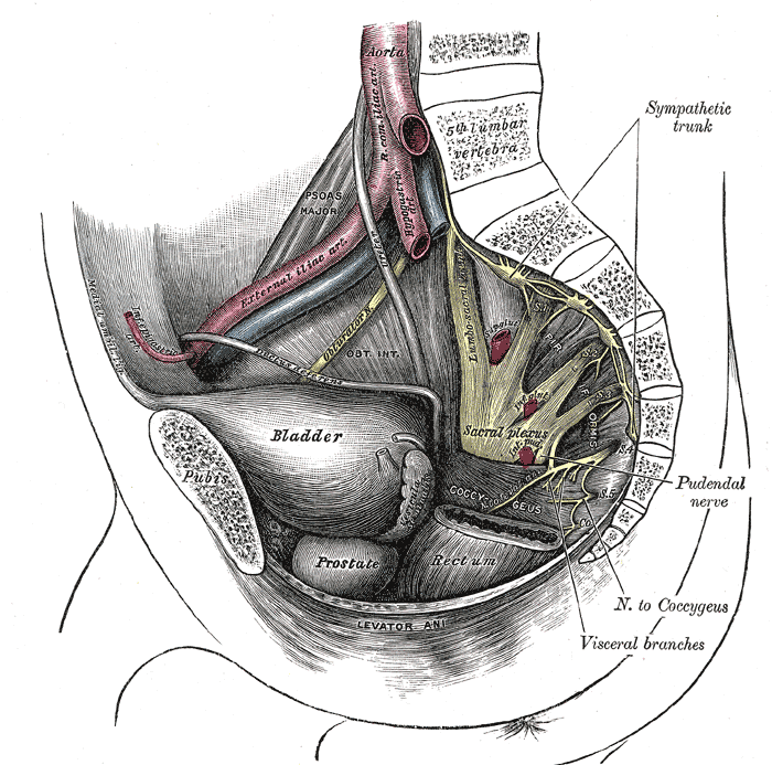
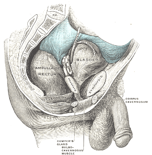 Male pelvic organs seen from right side.
 -
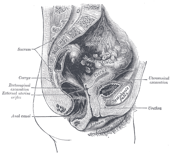 Median sagittal section of female pelvis.
 -
 The interior of bladder.
 -
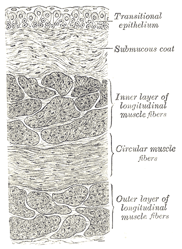 Vertical section of bladder wall.
 -
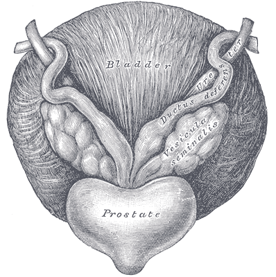 Fundus of the bladder with the vesiculæ seminales.
 -
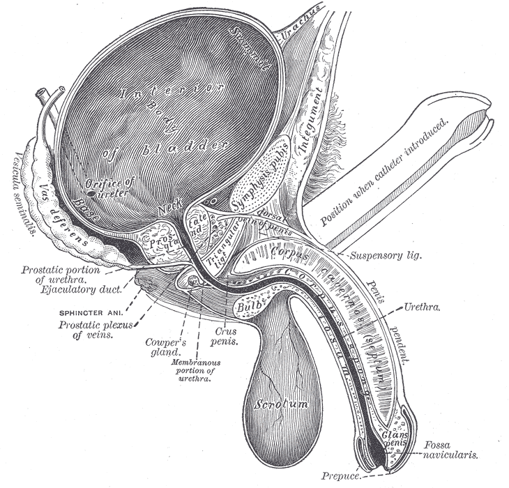 Vertical section of bladder, penis, and urethra.
 -
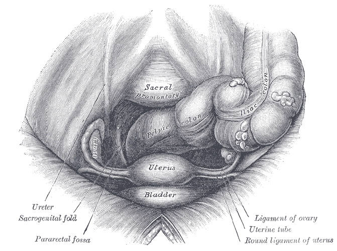 Female pelvis and its contents, seen from above and in front.
 -
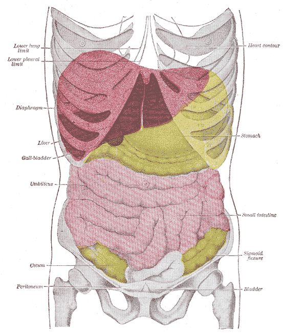 Topography of thoracic and abdominal viscera.
 -
The bladder can be seen highlighted in yellow in the illustration.
-
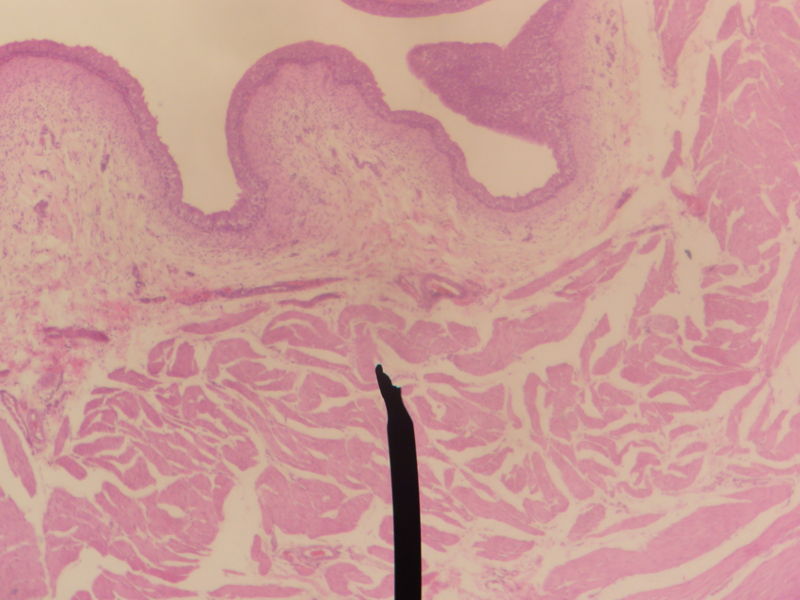 Layers of the urinary bladder wall and cross section of the detrusor muscle.


bg:Пикочен мехур cs:Močový měchýř cy:Pledren da:Urinblære de:Harnblase id:Kandung kemih he:שלפוחית השתן it:Vescica urinaria lt:Šlapimo pūslė nl:Urineblaas no:Urinblære sk:Močový mechúr fi:Virtsarakko sv:Urinblåsa yi:בלעדער
 EncycloReader
is supported by the
EncycloReader
is supported by the