MARCO
 From Wikipedia - Reading time: 9 min
Macrophage receptor with collagenous structure (MARCO) is a protein that in humans is encoded by the MARCO gene.[5][6][7][8] MARCO is a class A scavenger receptor that is found on particular subsets of macrophages.[9][10][11] Scavenger receptors are pattern recognition receptors (PRRs) found most commonly on immune cells.[10] Their defining feature is that they bind to polyanions and modified forms of a type of cholesterol called low-density lipoprotein (LDL).[9][10] MARCO is able to bind and phagocytose these ligands and pathogen-associated molecular patterns (PAMPs), leading to the clearance of pathogens and cell signaling events that lead to inflammation.[11][12] As part of the innate immune system, MARCO clears, or scavenges, pathogens, which leads to inflammatory responses.[12] The scavenger receptor cysteine-rich (SRCR) domain at the end of the extracellular side of MARCO binds ligands to activate the subsequent immune responses.[12] MARCO expression on macrophages has been associated with tumor development and also with Alzheimer's disease, via decreased responses of cells when ligands bind to MARCO.[13][14]
Structure
[edit]
MARCO is a transmembrane protein that has 5 domains (see figure).[10] The domain within the cell is called the cytoplasmic domain, as well as a transmembrane domain.[10] The extracellular regions of MARCO include a spacer domain, a collagenous domain, and the SRCR domain. The SRCR domain is required for MARCO binding to ligands, via 2 highly conserved arginine residues, termed the RxR motif.[10][12]
Other members of the class A scavenger receptors tend to have alpha helical coiled coil domains, but MARCO does not.[9] The C-terminal SRCR domain of MARCO affects the ability of the receptor to bind and take up ligand, activate inflammatory signaling, and adhere to surfaces.[12]
Cell expression
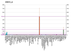
[edit]MARCO is expressed on a subset of tissue-resident macrophages in normal tissues, as well as circulating monocytes, dendritic cells, and B cells.[10][15] MARCO is typically present on the macrophages in the marginal zone of the spleen and the medullary lymph nodes, but it is also found in the liver.[13] Dendritic cells increase expression of MARCO when exposed to certain pathogens, resulting in alterations to the cytoskeleton of dendritic cells and increased phagocytosis.[9][16][10]
MARCO is highly expressed by macrophages and monocytes in the tumor microenvironment (TME), including tumor-associated macrophages (TAMs) and monocytic myeloid-derived suppressor cells (mMDSCs).[17][18] Expression of MARCO correlated (FDR < 0.01 and R > 0.2126212) with expression of genes associated with immunosuppressive TAMs, such as CD68, CD163, MSR1, IL4R, CHIA, TGFB1, IL10, and IL37, whereas no correlation was observed with expression of inducible nitric oxide synthase (NOS2), which is expressed by macrophages with an anti-tumor phenotype.
Functions
[edit]Phagocytosis
[edit]The primary function of scavenger receptors is to regulate phagocytosis of pathogens, but they also participate in cell–cell recognition and initiation of inflammatory responses.[11][10] MARCO, being a PRR, is able to bind to a wide variety of bacteria, making it an important receptor for activating an immune response against bacteria.[12] Soluble LPS and entire bacteria can each bind bind to MARCO,[19] as well as acetylated LDL (AcLDL), oxidized LDL (OxLDL), B cells in the marginal zone of the spleen, and apoptotic cells.[9][10] MARCO is therefore able to recognize and phagocytose pathogens and apoptotic cells, and operates independently of opsonization.[12]
Regulation of Inflammation
[edit]MARCO does not directly cause an inflammatory response, but it can interact with PAMPs to promote inflammation.[11][12] One way MARCO does this is by tethering a pathogen to other receptors on the cell, including PRRs such as TLR2,[12] which then lead to the activation of the transcription factor NF-κB, which regulates expression of genes that encode cytokines.[12] Through phagocytosis, MARCO also brings pathogens into the cell, which are processed by intracellular compartments that contain other signaling receptors such as TLR3, NOD2, and NALP3.[11] However, lung cancer cells polarize macrophages to express MARCO and acquire an immune-suppressive phenotype, through the release of IL37.[18] MARCO-expressing TAMs blocked activation of cytotoxic T cells and NK cells, inhibiting their proliferation, cytokine production, and tumor-cell killing capacity. Furthermore, MARCO+ macrophages increased proliferation of T-regulatory (Treg) cells and production of IL10, and diminished activity of CD8+ T cells.[18] Blocking MARCO or knocking out IL37 in lung cancer cell lines repolarized TAMs, resulting in recovered cytolytic activity and anti-tumor effects of natural killer (NK) cells and T cells, as well as reduced Treg-cell activities.[18]
Association with diseases
[edit]Alzheimer's disease
[edit]The activity of MARCO on microglia, the macrophages of the brain, is believed to be altered in development of Alzheimer's disease.[11][14] One primary characteristic of Alzheimer's disease is the presence of numerous senile plaques in the brain that contain amyloid beta peptides (Aβ).[14] Initially, the microglia clear the Aβ, which binds to receptors such as MARCO.[14] During development of Alzheimer's disease, however, the ability of microglia to clear Aβ is decreased, resulting in Aβ accumulation,[14] which is neurotoxic.[14] MARCO also interacts with formyl peptide receptor 2 (FPR2) to form a complex that causes the microglia to release inflammatory cytokines, which can also cause damage to neurons.[14]
Cancer
[edit]Antibodies against MARCO have been shown to slow growth and metastasis in syngeneic mouse tumor models, by reprogramming immunosuppressive (M2)-like TAMs into inflammatory (M1)-like macrophages. This switch involves changes to the metabolic program of the macrophages and activation of NK cells.[17][20] In addition, targeting MARCO on human macrophages repolarizes TAMs and restores the cytotoxic, anti-tumor capacities of NK and T cells.[18][20] These findings indicate that strategies that target MARCO-expressing TAMs to remodel the immune-suppressive tumor microenvironment might be developed for cancer therapy.
In non-small-cell lung cancer (NSCLC) tissues, researchers found an association between expression of MARCO mRNA and genes that regulate immune response pathways, including immunosuppressive TAMs, T-cell infiltration, and immune checkpoint molecules. Higher infiltration of tumor tissues by macrophages was seen in tumors expressing PD-L1; macrophages within tumor cell nests co-expressed MARCO and PD-L1. MARCO is therefore a target of immune therapeutic strategies to inhibit TAMs in NSCLC, possibly in combination with immune checkpoint inhibitors.[21]
In patients with renal cell carcinoma or colorectal cancer, those with tumors that expressed low levels of MARCO had longer survival times than patients whose tumors expressed high levels of MARCO. Additionally, high MARCO levels are observed in patients with melanoma that is refractory to checkpoint inhibitor therapy, and patients with solid tumors refractory to chemotherapy.[22]
References
[edit]- ^ a b c GRCh38: Ensembl release 89: ENSG00000019169 – Ensembl, May 2017
- ^ a b c GRCm38: Ensembl release 89: ENSMUSG00000026390 – Ensembl, May 2017
- ^ "Human PubMed Reference:". National Center for Biotechnology Information, U.S. National Library of Medicine.
- ^ "Mouse PubMed Reference:". National Center for Biotechnology Information, U.S. National Library of Medicine.
- ^ Elomaa O, Sankala M, Pikkarainen T, Bergmann U, Tuuttila A, Raatikainen-Ahokas A, et al. (February 1998). "Structure of the human macrophage MARCO receptor and characterization of its bacteria-binding region". The Journal of Biological Chemistry. 273 (8): 4530–4538. doi:10.1074/jbc.273.8.4530. PMID 9468508.
- ^ Elomaa O, Kangas M, Sahlberg C, Tuukkanen J, Sormunen R, Liakka A, et al. (February 1995). "Cloning of a novel bacteria-binding receptor structurally related to scavenger receptors and expressed in a subset of macrophages". Cell. 80 (4): 603–609. doi:10.1016/0092-8674(95)90514-6. PMID 7867067. S2CID 18632239.
- ^ Kangas M, Brännström A, Elomaa O, Matsuda Y, Eddy R, Shows TB, Tryggvason K (May 1999). "Structure and chromosomal localization of the human and murine genes for the macrophage MARCO receptor". Genomics. 58 (1): 82–89. doi:10.1006/geno.1999.5811. PMID 10331948.
- ^ "Entrez Gene: MARCO macrophage receptor with collagenous structure".
- ^ a b c d e Plüddemann A, Neyen C, Gordon S (November 2007). "Macrophage scavenger receptors and host-derived ligands". Methods. 43 (3): 207–217. doi:10.1016/j.ymeth.2007.06.004. PMID 17920517.
- ^ a b c d e f g h i j Bowdish DM, Gordon S (January 2009). "Conserved domains of the class A scavenger receptors: evolution and function". Immunological Reviews. 227 (1): 19–31. doi:10.1111/j.1600-065x.2008.00728.x. PMID 19120472. S2CID 10481905.
- ^ a b c d e f Mukhopadhyay S, Varin A, Chen Y, Liu B, Tryggvason K, Gordon S (January 2011). "SR-A/MARCO-mediated ligand delivery enhances intracellular TLR and NLR function, but ligand scavenging from cell surface limits TLR4 response to pathogens". Blood. 117 (4): 1319–1328. doi:10.1182/blood-2010-03-276733. PMID 21098741. S2CID 206890578.
- ^ a b c d e f g h i j Novakowski KE, Huynh A, Han S, Dorrington MG, Yin C, Tu Z, et al. (August 2016). "A naturally occurring transcript variant of MARCO reveals the SRCR domain is critical for function". Immunology and Cell Biology. 94 (7): 646–655. doi:10.1038/icb.2016.20. PMC 4980223. PMID 26888252.
- ^ a b Sun H, Song J, Weng C, Xu J, Huang M, Huang Q, et al. (May 2017). "Association of decreased expression of the macrophage scavenger receptor MARCO with tumor progression and poor prognosis in human hepatocellular carcinoma". Journal of Gastroenterology and Hepatology. 32 (5): 1107–1114. doi:10.1111/jgh.13633. PMID 27806438. S2CID 9564859.
- ^ a b c d e f g Yu Y, Ye RD (January 2015). "Microglial Aβ receptors in Alzheimer's disease". Cellular and Molecular Neurobiology. 35 (1): 71–83. doi:10.1007/s10571-014-0101-6. PMC 11486233. PMID 25149075. S2CID 15704092.
- ^ Getts DR, Terry RL, Getts MT, Deffrasnes C, Müller M, van Vreden C, et al. (January 2014). "Therapeutic inflammatory monocyte modulation using immune-modifying microparticles". Science Translational Medicine. 6 (219): 219ra7. doi:10.1126/scitranslmed.3007563. PMC 3973033. PMID 24431111.
- ^ Kissick HT, Dunn LK, Ghosh S, Nechama M, Kobzik L, Arredouani MS (2014-08-04). "The scavenger receptor MARCO modulates TLR-induced responses in dendritic cells". PLOS ONE. 9 (8): e104148. Bibcode:2014PLoSO...9j4148K. doi:10.1371/journal.pone.0104148. PMC 4121322. PMID 25089703.
- ^ a b Georgoudaki AM, Prokopec KE, Boura VF, Hellqvist E, Sohn S, Östling J, et al. (May 2016). "Reprogramming Tumor-Associated Macrophages by Antibody Targeting Inhibits Cancer Progression and Metastasis". Cell Reports. 15 (9): 2000–2011. doi:10.1016/j.celrep.2016.04.084. PMID 27210762.
- ^ a b c d e La Fleur L, Botling J, He F, Pelicano C, Zhou C, He C, et al. (February 2021). "Targeting MARCO and IL37R on Immunosuppressive Macrophages in Lung Cancer Blocks Regulatory T Cells and Supports Cytotoxic Lymphocyte Function". Cancer Research. 81 (4): 956–967. doi:10.1158/0008-5472.CAN-20-1885. PMID 33293426. S2CID 228079815.
- ^ Athanasou NA (May 2016). "The pathobiology and pathology of aseptic implant failure". Bone & Joint Research. 5 (5): 162–168. doi:10.1302/2046-3758.55.bjr-2016-0086. PMC 4921050. PMID 27146314.
- ^ a b Eisinger S, Sarhan D, Boura VF, Ibarlucea-Benitez I, Tyystjärvi S, Oliynyk G, et al. (December 2020). "Targeting a scavenger receptor on tumor-associated macrophages activates tumor cell killing by natural killer cells". Proceedings of the National Academy of Sciences of the United States of America. 117 (50): 32005–32016. Bibcode:2020PNAS..11732005E. doi:10.1073/pnas.2015343117. PMC 7750482. PMID 33229588.
- ^ La Fleur L, Boura VF, Alexeyenko A, Berglund A, Pontén V, Mattsson JS, et al. (October 2018). "Expression of scavenger receptor MARCO defines a targetable tumor-associated macrophage subset in non-small cell lung cancer". International Journal of Cancer. 143 (7): 1741–1752. doi:10.1002/ijc.31545. PMID 29667169. S2CID 4941508.
- ^ Jahchan NS, Mujal AM, Pollack JL, Binnewies M, Sriram V, Reyno L, Krummel MF (2019). "Tuning the Tumor Myeloid Microenvironment to Fight Cancer". Frontiers in Immunology. 10: 1611. doi:10.3389/fimmu.2019.01611. PMC 6673698. PMID 31402908.
Further reading
[edit]- Elshourbagy NA, Li X, Terrett J, Vanhorn S, Gross MS, Adamou JE, et al. (February 2000). "Molecular characterization of a human scavenger receptor, human MARCO". European Journal of Biochemistry. 267 (3): 919–926. doi:10.1046/j.1432-1327.2000.01077.x. PMID 10651831.
- Seta N, Granfors K, Sahly H, Kuipers JG, Song YW, Baeten D, et al. (April 2001). "Expression of host defense scavenger receptors in spondylarthropathy". Arthritis and Rheumatism. 44 (4): 931–939. doi:10.1002/1529-0131(200104)44:4<931::AID-ANR150>3.0.CO;2-T. PMID 11315932.
- Brännström A, Sankala M, Tryggvason K, Pikkarainen T (February 2002). "Arginine residues in domain V have a central role for bacteria-binding activity of macrophage scavenger receptor MARCO". Biochemical and Biophysical Research Communications. 290 (5): 1462–1469. doi:10.1006/bbrc.2002.6378. PMID 11820786.
- Sankala M, Brännström A, Schulthess T, Bergmann U, Morgunova E, Engel J, et al. (September 2002). "Characterization of recombinant soluble macrophage scavenger receptor MARCO". The Journal of Biological Chemistry. 277 (36): 33378–33385. doi:10.1074/jbc.M204494200. PMID 12097327.
- Mirani M, Elenkov I, Volpi S, Hiroi N, Chrousos GP, Kino T (December 2002). "HIV-1 protein Vpr suppresses IL-12 production from human monocytes by enhancing glucocorticoid action: potential implications of Vpr coactivator activity for the innate and cellular immunity deficits observed in HIV-1 infection". Journal of Immunology. 169 (11): 6361–6368. doi:10.4049/jimmunol.169.11.6361. PMID 12444143.
- Bin LH, Nielson LD, Liu X, Mason RJ, Shu HB (July 2003). "Identification of uteroglobin-related protein 1 and macrophage scavenger receptor with collagenous structure as a lung-specific ligand-receptor pair". Journal of Immunology. 171 (2): 924–930. doi:10.4049/jimmunol.171.2.924. PMID 12847263.
- Arredouani MS, Palecanda A, Koziel H, Huang YC, Imrich A, Sulahian TH, et al. (November 2005). "MARCO is the major binding receptor for unopsonized particles and bacteria on human alveolar macrophages". Journal of Immunology. 175 (9): 6058–6064. doi:10.4049/jimmunol.175.9.6058. PMID 16237101.
- Liu T, Qian WJ, Gritsenko MA, Camp DG, Monroe ME, Moore RJ, Smith RD (2006). "Human plasma N-glycoproteome analysis by immunoaffinity subtraction, hydrazide chemistry, and mass spectrometry". Journal of Proteome Research. 4 (6): 2070–2080. doi:10.1021/pr0502065. PMC 1850943. PMID 16335952.
 KSF
KSF


