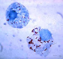
Rickettsia
 From Wikipedia - Reading time: 13 min
Rickettsia is a genus of nonmotile, gram-negative, nonspore-forming, highly pleomorphic bacteria that may occur in the forms of cocci (0.1 μm in diameter), bacilli (1–4 μm long), or threads (up to about 10 μm long). The genus was named after Howard Taylor Ricketts in honor of his pioneering work on tick-borne spotted fever.
Properly, Rickettsia is the name of a single genus, but the informal term "rickettsia", plural "rickettsias," usually not capitalised, commonly applies to any members of the order Rickettsiales. Being obligate intracellular bacteria, rickettsias depend on entry, growth, and replication within the cytoplasm of living eukaryotic host cells (typically endothelial cells).[9] Accordingly, Rickettsia species cannot grow in artificial nutrient culture; they must be grown either in tissue or embryo cultures. Mostly chicken embryos are used, following a method developed by Ernest William Goodpasture and his colleagues at Vanderbilt University in the early 1930s. Many new strains or species of Rickettsia are described each year.[10][11] Some Rickettsia species are pathogens of medical and veterinary interest, but many Rickettsia are non-pathogenic to vertebrates, including humans, and infect only arthropods, often non-hematophagous, such as aphids or whiteflies.[12][13][14] Many Rickettsia species are thus arthropod-specific symbionts, but are often confused with pathogenic Rickettsia (especially in medical literature), showing that the current view in rickettsiology has a strong anthropocentric bias.[15]
Pathogenic Rickettsia species are transmitted by numerous types of arthropods, including chiggers, ticks, fleas, and lice, and are associated with both human and plant diseases.[16] Most notably, Rickettsia species are the pathogens responsible for typhus, rickettsialpox, boutonneuse fever, African tick-bite fever, Rocky Mountain spotted fever, Flinders Island spotted fever, and Queensland tick typhus (Australian tick typhus).[17] The majority of pathogenic Rickettsia bacteria are susceptible to antibiotics of the tetracycline group.
Classification
[edit]The classification of Rickettsia into three groups (spotted fever, typhus, and scrub typhus) was initially based on serology. This grouping has since been confirmed by DNA sequencing. All three of these groups include human pathogens. The scrub typhus group has been reclassified as a related new genus, Orientia, but they still are in the order Rickettsiales and accordingly still are grouped with the rest of the rickettsial diseases.[citation needed]
Rickettsias are more widespread than previously believed and are known to be associated with arthropods, leeches, and protists. Divisions have also been identified in the spotted fever group and this group likely should be divided into two clades.[18] Arthropod-inhabiting rickettsiae are generally associated with reproductive manipulation (such as parthenogenesis) to persist in host lineage.[16]
In March 2010, Swedish researchers reported a case of bacterial meningitis in a woman caused by Rickettsia helvetica previously thought to be harmless.[19]
Spotted fever group
[edit]- Rickettsia rickettsii (Western Hemisphere)
- Rickettsia akari (USA, former Soviet Union)
- Rickettsia conorii (Mediterranean countries, Africa, Southwest Asia, India)
- Rickettsia sibirica (Siberia, Mongolia, northern China)
- Rickettsia australis (Australia)
- Rickettsia felis (North and South America, Southern Europe, Australia)
- Rickettsia africae (South Africa)
- Rickettsia hoogstraalii (Croatia, Spain and Georgia USA)[20]
- Unknown pathogenicity
Typhus group
[edit]- Rickettsia prowazekii (worldwide)
- Epidemic typhus, recrudescent typhus, and sporadic typhus
- Rickettsia typhi (worldwide)
- Murine typhus (endemic typhus)
Scrub typhus group
[edit]- The causative agent of scrub typhus formerly known as R. tsutsugamushi has been reclassified into the genus Orientia.
| Schematic ribosomal RNA phylogeny of Alphaproteobacteria | ||||||||||||||||||||||||
| ||||||||||||||||||||||||
| The cladogram of Rickettsidae has been inferred by Ferla et al. [21] from the comparison of 16S + 23S ribosomal RNA sequences. |
Flora and fauna pathogenesis
[edit]Plant diseases have been associated with these Rickettsia-like organisms (RLOs):[22]
- Beet latent rosette RLO
- Citrus greening bacterium possibly this citrus greening disease
- Clover leaf RLO
- Grapevine infectious necrosis RLO
- Grapevine Pierce's RLO
- Grapevine yellows RLO
- Witch's broom disease on Larix spp.
- Peach phony RLO
- Papaya Bunchy Top Disease[23]
Infection occurs in nonhuman mammals; for example, species of Rickettsia have been found to afflict the South American guanaco, Lama guanacoe[24] potentially marsupials[25][26] and reptiles.[27]
Pathophysiology
[edit]This section needs expansion. You can help by adding to it. (August 2013) |
Rickettsial organisms are obligate intracellular parasites and invade vascular endothelial cells in target organs, damaging them and producing increased vascular permeability with consequent oedema, hypotension, and hypoalbuminaemia.[28]
Genomics
[edit]Certain segments of rickettsial genomes resemble those of mitochondria.[29] The deciphered genome of R. prowazekii is 1,111,523 bp long and contains 834 genes.[30] Unlike free-living bacteria, it contains no genes for anaerobic glycolysis or genes involved in the biosynthesis and regulation of amino acids and nucleosides. In this regard, it is similar to mitochondrial genomes; in both cases, nuclear (host) resources are used.
ATP production in Rickettsia is the same as that in mitochondria. In fact, of all the microbes known, the Rickettsia is probably the closest relative (in a phylogenetic sense) to the mitochondria. Unlike the latter, the genome of R. prowazekii, however, contains a complete set of genes encoding for the tricarboxylic acid cycle and the respiratory chain complex. Still, the genomes of the Rickettsia, as well as the mitochondria, are frequently said to be "small, highly derived products of several types of reductive evolution".
The recent discovery of another parallel between Rickettsia and viruses may become a basis for fighting HIV infection.[31] Human immune response to the scrub typhus pathogen, Orientia tsutsugamushi, appears to provide a beneficial effect against HIV infection progress, negatively influencing the virus replication process. A probable reason for this actively studied phenomenon is a certain degree of homology between the rickettsiae and the virus, namely, common epitope(s) due to common genome fragment(s) in both pathogens. Surprisingly, the other infection reported to be likely to provide the same effect (decrease in viral load) is the virus-caused illness dengue fever.
Comparative analysis of genomic sequences have also identified five conserved signature indels in important proteins, which are uniquely found in members of the genus Rickettsia. These indels consist of a four-amino-acid insertion in transcription repair coupling factor Mfd, a 10-amino-acid insertion in ribosomal protein L19, a one-amino-acid insertion in FtsZ, a one-amino-acid insertion in major sigma factor 70, and a one-amino-acid deletion in exonuclease VII. These indels are all characteristic of the genus and serve as molecular markers for Rickettsia.[32]
Bacterial small RNAs play critical roles in virulence and stress/adaptation responses. Although their specific functions have not been discovered in Rickettsia, few studies showed the expression of novel sRNA in human microvascular endothelial cells (HMEC) infected with Rickettsia.[33][34]
Genomes of intracellular or parasitic bacteria undergo massive reduction compared to their free-living relatives. Examples include Rickettsia for alpha proteobacteria, T. whipplei for Actinobacteria, Mycoplasma for Firmicutes (the low G+C content Gram-positive), and Wigglesworthia and Buchnera for gamma proteobacteria.[35]
Naming
[edit]The genus Rickettsia is named after Howard Taylor Ricketts (1871–1910), who studied Rocky Mountain spotted fever in the Bitterroot Valley of Montana, and eventually died of typhus after studying that disease in Mexico City.
In his early part of career, he undertook research at Northwestern University on blastomycosis. He later worked on Rocky Mountain spotted fever at the University of Chicago and Bitterroot Valley of Montana. He was so devoted to his research that on several occasions, he injected himself with pathogens to study their effects. On account of the apparent similarity between Rocky Mountain fever and typhus fever, he became occupied in investigating the latter in Chicago where the disease was epidemic, and became a victim of the epidemic in 1910. His investigations and discoveries added materially to the sum of medical knowledge.
References
[edit]- ^ a b c d e f g h Skerman VB, McGowan V, Sneath PH, eds. (1989). Approved Lists of Bacterial Names (amended ed.). Washington, DC: American Society for Microbiology.
- ^ Truper HG, De' Clari L (1997). "Taxonomic note: Necessary correction of specific epithets formed as substantives (nouns) 'in apposition'". Int J Syst Bacteriol. 47 (3): 908–909. doi:10.1099/00207713-47-3-908.
- ^ Beati, L.; Meskini, M., et al. (1997), "Rickettsia aeschlimannii sp. nov., a new spotted fever group rickettsia associated with Hyalomma marginatum ticks", Int J Syst Bacteriol 47 (2): 548-55s4
- ^ Kelly PJ, Beati L, Mason PR, Matthewman LA, Roux V, Raoult D (April 1996). "Rickettsia africae sp. nov., the etiological agent of African tick bite fever". International Journal of Systematic Bacteriology. 46 (2): 611–614. doi:10.1099/00207713-46-2-611. PMID 8934912.
- ^ Fujita, H.; Fournier, P.-E., et al. (2006), "Rickettsia asiatica sp. nov., isolated in Japan", Int J Syst Evol Microbiol 56 (Pt 10): 2365–2368
- ^ Billings AN, Teltow GJ, Weaver SC, Walker DH (1998). "Molecular characterization of a novel Rickettsia species from Ixodes scapularis in Texas" (PDF). Emerging Infectious Diseases. 4 (2): 305–309. doi:10.3201/eid0402.980221. PMC 2640119. PMID 9621204. Archived from the original (PDF) on 8 August 2017.
- ^ La Scola, B.; Meconi, S., et al. (2002), "Emended description of Rickettsia felis (Bouyer et al. 2001), a temperature-dependent cultured bacterium"[permanent dead link], Int J Syst Evol Microbiol 52 (Pt 6): 2035–2041
- ^ "Rickettsia". NCBI taxonomy. Bethesda, MD: National Center for Biotechnology Information. Retrieved 8 January 2019.
- ^ Walker DH (1996). Baron S, et al. (eds.). Rickettsiae. In: Barron's Medical Microbiology (4th ed.). Univ of Texas Medical Branch. ISBN 978-0-9631172-1-2. (via NCBI Bookshelf).
- ^ Binetruy F, Buysse M, Barosi R, Duron O (February 2020). "Novel Rickettsia genotypes in ticks in French Guiana, South America". Scientific Reports. 10 (1): 2537. Bibcode:2020NatSR..10.2537B. doi:10.1038/s41598-020-59488-0. PMC 7018960. PMID 32054909.
- ^ Buysse M, Duron O (May 2020). "Two novel Rickettsia species of soft ticks in North Africa: 'Candidatus Rickettsia africaseptentrionalis' and 'Candidatus Rickettsia mauretanica'". Ticks and Tick-Borne Diseases. 11 (3): 101376. doi:10.1016/j.ttbdis.2020.101376. PMID 32005627. S2CID 210997920.
- ^ Sakurai M, Koga R, Tsuchida T, Meng XY, Fukatsu T (July 2005). "Rickettsia symbiont in the pea aphid Acyrthosiphon pisum: novel cellular tropism, effect on host fitness, and interaction with the essential symbiont Buchnera". Applied and Environmental Microbiology. 71 (7): 4069–4075. Bibcode:2005ApEnM..71.4069S. doi:10.1128/AEM.71.7.4069-4075.2005. PMC 1168972. PMID 16000822.
- ^ Himler AG, Adachi-Hagimori T, Bergen JE, Kozuch A, Kelly SE, Tabashnik BE, et al. (April 2011). "Rapid spread of a bacterial symbiont in an invasive whitefly is driven by fitness benefits and female bias". Science. 332 (6026): 254–256. Bibcode:2011Sci...332..254H. doi:10.1126/science.1199410. PMID 21474763. S2CID 31371994.
- ^ Giorgini M, Bernardo U, Monti MM, Nappo AG, Gebiola M (April 2010). "Rickettsia symbionts cause parthenogenetic reproduction in the parasitoid wasp Pnigalio soemius (Hymenoptera: Eulophidae)". Applied and Environmental Microbiology. 76 (8): 2589–2599. Bibcode:2010ApEnM..76.2589G. doi:10.1128/AEM.03154-09. PMC 2849191. PMID 20173065.
- ^ Labruna MB, Walker DH (October 2014). "Rickettsia felis and changing paradigms about pathogenic rickettsiae". Emerging Infectious Diseases. 20 (10): 1768–1769. doi:10.3201/eid2010.131797. PMC 4193273. PMID 25271441.
- ^ a b Perlman SJ, Hunter MS, Zchori-Fein E (September 2006). "The emerging diversity of Rickettsia". Proceedings. Biological Sciences. 273 (1598): 2097–2106. doi:10.1098/rspb.2006.3541. PMC 1635513. PMID 16901827.
- ^ Unsworth NB, Stenos J, Graves SR, Faa AG, Cox GE, Dyer JR, et al. (April 2007). "Flinders Island spotted fever rickettsioses caused by "marmionii" strain of Rickettsia honei, Eastern Australia". Emerging Infectious Diseases. 13 (4): 566–573. doi:10.3201/eid1304.050087. PMC 2725950. PMID 17553271.
- ^ Gillespie JJ, Beier MS, Rahman MS, Ammerman NC, Shallom JM, Purkayastha A, et al. (March 2007). "Plasmids and rickettsial evolution: insight from Rickettsia felis". PLOS ONE. 2 (3): e266. Bibcode:2007PLoSO...2..266G. doi:10.1371/journal.pone.0000266. PMC 1800911. PMID 17342200.
 .
.
- ^ "Rickettsia helvetica in Patient with Meningitis, Sweden, 2006" Emerging Infectious Diseases, Volume 16, Number 3 – March 2010
- ^ Duh, D., V. Punda-Polic, T. Avsic-Zupanc, D. Bouyer, D.H. Walker, V.L. Popov, M. Jelovsek, M. Gracner, T. Trilar, N. Bradaric, T.J. Kurtti and J. Strus. (2010) Rickettsia hoogstraalii sp. nov., isolated from hard- and soft-bodied ticks. International Journal of Systematic and Evolutionary Microbiology, 60, 977–984; [1], accessed 16 July 2010.
- ^ Ferla MP, Thrash JC, Giovannoni SJ, Patrick WM (2013). "New rRNA gene-based phylogenies of the Alphaproteobacteria provide perspective on major groups, mitochondrial ancestry and phylogenetic instability". PLOS ONE. 8 (12): e83383. Bibcode:2013PLoSO...883383F. doi:10.1371/journal.pone.0083383. PMC 3859672. PMID 24349502.
- ^ Smith IM, Dunez J, Lelliot RA, Phillips DH, Archer SA (1988). European Handbook of Plant Diseases. Blackwell Scientific Publications. ISBN 978-0-632-01222-0.
- ^ Davis, M. J. 1996
- ^ C. Michael Hogan. 2008. Guanaco: Lama guanicoe, GlobalTwitcher.com, ed. N. Strömberg Archived 4 March 2011 at the Wayback Machine
- ^ Vilcins IE, Old JM, Deane EM (2009). Molecular detection of Rickettsia, Coxiella and Rickettsiella in three Australian native tick species. Experimental and Applied Acarology. 49(3), 229-242. DOI: 10.1007/s10493-009-9260-4
- ^ Vilcins IE, Old JM, Deane EM (2008). Detection of a spotted fever group Rickettsia in the tick Ixodes tasmani collected from koalas (Phascolarctos cinereus) in Port Macquarie, N.S.W. Journal of Medical Entomology. 45(4), 745-750. DOI: 10.1016/j.vetpar.2009.02.015
- ^ Vilcins I, Fournier P, Old JM, Deane EM (2009). Evidence for the presence of Francisella and spotted fever group Rickettsia DNA in the Tick Amblyomma fimbriatum (Acari: Ixodidae), Northern Territory, Australia. Journal of Medical Entomology. 46(4), 926-933. doi: 10.1186/s13071-015-0719-3
- ^ Rathore MH (14 June 2016). "Rickettsial infection: Overview". Medscape. Retrieved 16 November 2017.
- ^ Emelyanov VV (April 2003). "Mitochondrial connection to the origin of the eukaryotic cell". European Journal of Biochemistry. 270 (8): 1599–1618. doi:10.1046/j.1432-1033.2003.03499.x. PMID 12694174.
- ^ Andersson SG, Zomorodipour A, Andersson JO, Sicheritz-Pontén T, Alsmark UC, Podowski RM, et al. (November 1998). "The genome sequence of Rickettsia prowazekii and the origin of mitochondria". Nature. 396 (6707): 133–140. Bibcode:1998Natur.396..133A. doi:10.1038/24094. PMID 9823893.
- ^ Kannangara S, DeSimone JA, Pomerantz RJ (September 2005). "Attenuation of HIV-1 infection by other microbial agents". The Journal of Infectious Diseases. 192 (6): 1003–1009. doi:10.1086/432767. PMID 16107952.
- ^ Gupta RS (January 2005). "Protein signatures distinctive of alpha proteobacteria and its subgroups and a model for alpha-proteobacterial evolution". Critical Reviews in Microbiology. 31 (2): 101–135. doi:10.1080/10408410590922393. PMID 15986834. S2CID 30170035.
- ^ Schroeder CL, Narra HP, Rojas M, Sahni A, Patel J, Khanipov K, et al. (December 2015). "Bacterial small RNAs in the Genus Rickettsia". BMC Genomics. 16: 1075. doi:10.1186/s12864-015-2293-7. PMC 4683814. PMID 26679185.
- ^ Schroeder CL, Narra HP, Sahni A, Rojas M, Khanipov K, Patel J, et al. (2016). "Identification and Characterization of Novel Small RNAs in Rickettsia prowazekii". Frontiers in Microbiology. 7: 859. doi:10.3389/fmicb.2016.00859. PMC 4896933. PMID 27375581.
- ^ Raoult D, Ogata H, Audic S, Robert C, Suhre K, Drancourt M, Claverie JM (August 2003). "Tropheryma whipplei Twist: a human pathogenic Actinobacteria with a reduced genome". Genome Research. 13 (8): 1800–1809. doi:10.1101/gr.1474603. PMC 403771. PMID 12902375.
External links
[edit]- Rickettsia genomes and related information at PATRIC, a Bioinformatics Resource Center funded by NIAID
- African Tick Bite Fever from the Centers for Disease Control and Prevention
 KSF
KSF