Bone
 From Nwe
 Bones are semi-rigid, porous, mineralized organs, consisting of cells in a hard matrix, that form part of the endoskeleton of vertebrates. Bones function to move, support, and protect the body, produce red and white blood cells, and store minerals.
Although externally bones may appear to be simple and even solid structures, in reality they are composed of living bone tissue interlaced with blood vessels, nerve fibers, and so forth, and their formation, structure, and function involves stunningly complex coordination.
Bones come in a variety of shapes and have a intricate internal and external structure, allowing them to be lightweight yet strong and hard, while fulfilling their many other functions. One of the types of tissues that make up bones is the mineralized osseous tissue, also called bone tissue, a specialized connective tissue that gives bones their rigidity and honeycomb-like, three-dimensional internal structure. Other tissue types found in bones in their entirety include marrow, the periosteum, nerves, blood vessels, and cartilage.
Because a group of tissues are involved that perform a specific function or group of functions, bones can be referred to as organs, although bone tissue is the dominant tissue, leading bone to often be classified as a specialized type of connective tissue.
Characteristics of bone
All bones consist of living cells embedded in the mineralized organic matrix that makes up the osseous tissue.
The primary tissue of bone, osseous tissue, is a relatively hard and lightweight composite material, formed mostly of calcium phosphate in the chemical arrangement termed calcium hydroxylapatite (this is the osseous tissue that gives bones their rigidity). It has relatively high compressive strength but poor tensile strength, meaning it resists pushing forces well, but not pulling forces. While bone is essentially brittle, it does have a significant degree of elasticity, contributed chiefly by collagen. Collagen is the main protein of connective tissue in animals and involves the harmonization of three polypeptide chains into the form of a triple helix. It is characterized by the regular arrangement of amino acids in each of the three chains; under tension, the triple helix coils tight, resisting stretching, and making collagen valuable for structure and support, while giving bones some elasticity.
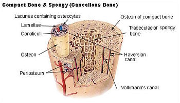
Bone is not a uniformly solid material, but rather has some spaces between its hard components. The hard outer layer of bones is called compact bone tissue due to its minimal gaps or spaces. This tissue gives bones their smooth, white, and solid appearance, and accounts for 80 percent of the total bone mass of an adult skeleton. Compact bone may also be referred to as dense bone or cortical bone. Filling the interior of the organ is the hole-filled spongy bone tissue (also called cancellous bone or trabecular bone) which is comprised of a network of flat or needle-shaped trabeculae, which makes the overall organ lighter and allows room for blood vessels and marrow. Spongy bone accounts for the remaining 20 percent of total bone mass, but has nearly ten times the surface area of compact bone.
The exterior of bones (except where they interact with other bones through joints) is covered by the periosteum, which has an external fibrous layer, and an internal osteogenic layer. The periosteum is richly supplied with blood, lymph, and nerve vessels, attaching to the bone itself through Sharpey's fibers.
Bone can also be either woven or lamellar (layered). Woven bone is weak, with a small number of randomly oriented collagen fibers, but forms quickly and without a preexisting structure during periods of repair or growth. Lamellar bone is stronger, formed of numerous stacked layers and filled with many collagen fibers parallel to other fibers in the same layer. The fibers run in opposite directions in alternating layers, assisting in the bone's ability to resist torsion forces. After a break, woven bone quickly forms and is gradually replaced by slow-growing lamellar bone on preexisting, calcified hyaline cartilage through a process known as "bony substitution."
Seven functions of bones
There are seven main functions of bones.
- Protection: Bones can serve to protect internal organs, such as the skull protects the brain or the ribs protect the abdomen.
- Shape: Bones provide a frame to keep the body supported.
- Blood production: The bone marrow, located within the medullary cavity of long bones and the interstices of cancellous bone, produces blood cells in a process called haematopoiesis.
- Mineral storage: Bones act as reserves of minerals important for the body, most notably calcium and phosphorus.
- Movement: Bones, skeletal muscles, tendons, ligaments, and joints function together to generate and transfer forces so that individual body parts or the whole body can be manipulated in three-dimensional space. The interaction between bone and muscle is studied in biomechanics.
- Acid-base balance: Bone buffers the blood against excessive pH changes by absorbing or releasing alkaline salts.
- Detoxification: Bone tissue removes heavy metals and other foreign elements from the blood and thus reduces their effects on nervous and other tissues. It can later release these more slowly for excretion.
Most bones perform all of these functions to one degree or another, but certain bones are more specialized for certain functions.
Five types of bones
There are five types of bones in the human body: long, short, flat, irregular, and sesamoid.
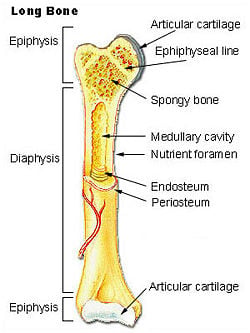
- Long bones are longer than they are wide, consisting of a long shaft (the diaphysis) plus two articular (joint) surfaces, called epiphyses. They are comprised mostly of compact bone, but are generally thick enough to contain considerable spongy bone and marrow in the hollow center (the medullary cavity). Most bones of the limbs (including the three bones of the fingers) are long bones, except for the kneecap (patella), and the carpal, metacarpal, tarsal, and metatarsal bones of the wrist and ankle. The classification refers to shape rather than the size.
- Short bones are roughly cube-shaped, and have only a thin layer of compact bone surrounding a spongy interior. The bones of the wrist and ankle are short bones, as are the sesamoid bones.
- Flat bones are thin and generally curved, with two parallel layers of compact bones sandwiching a layer of spongy bone. Most of the bones of the skull are flat bones, as is the sternum.
- Irregular bones do not fit into the above categories. They consist of thin layers of compact bone surrounding a spongy interior. As implied by the name, their shapes are irregular and complicated. The bones of the spine and hips are irregular bones.
- Sesamoid bones are short bones embedded in tendons. Since they act to hold the tendon further away from the joint, the angle of the tendon is increased and thus the force of the muscle is increased. Examples of sesamoid bones are the patella and the pisiform.
Bone cells
- Osteoblasts are mononucleate bone-forming cells which descend from osteoprogenitor cells. They are located on the surface of osteoid seams and make a protein mixture known as osteoid, which mineralizes to becomes bone. Osteoid is primarily composed of Type I collagen and manufactures hormones, such as prostaglandins, to act on the bone itself. They robustly produce alkaline phosphatase, an enzyme that has a role in the mineralization of bone, as well as many matrix proteins. Osteoblasts are the immature bone cells.
- Bone lining cells are essentially inactive osteoblasts. They cover all of the available bone surface and function as a barrier for certain ions.
- Osteocytes originate from osteoblasts, which have migrated into and become trapped and surrounded by bone matrix which they themselves produce. The spaces which they occupy are known as lacunae. Osteocytes have many processes which reach out to meet osteoblasts probably for the purposes of communication. Their functions include to varying degrees: formation of bone, matrix maintenance and calcium homeostasis. They possibly act as mechano-sensory receptors—regulating the bone's response to stress. They are mature bone cells.
- Osteoclasts are the cells responsible for bone resorption (remodeling of bone to reduce its volume). Osteoclasts are large, multinucleated cells located on bone surfaces in what are called Howship's lacunae or resorption pits. These lacunae, or resorption pits, are left behind after the breakdown of bone and often present as scalloped surfaces. Because the osteoclasts are derived from a monocyte stem-cell lineage, they are equipped with engulfment strategies similar to circulating macrophages. Osteoclasts mature and/or migrate to discrete bone surfaces. Upon arrival, active enzymes, such as tartrate resistant acid phosphatase, are secreted against the mineral substrate.
The process of bone resorption releases stored calcium into the systemic circulation and is an important process in regulating calcium balance. As bone formation actively fixes circulating calcium in its mineral form, removing it from the bloodstream, resorption actively unfixes it, thereby increasing circulating calcium levels. These processes occur in tandem at site-specific locations and are known as bone turnover or remodeling. Osteoblasts and osteoclasts, coupled together via paracrine cell signaling, are referred to as bone remodeling units. The iteration of remodeling events at the cellular level is influential on shaping and sculpting the skeleton during growth and in response to stress (such as weight-bearing exercise or bone healing).
Matrix
The matrix comprises the other major constituent of bone. It has inorganic and organic parts. The inorganic is mainly crystalline mineral salts and calcium, which is present in the form of hydroxyapatite. The matrix is initially laid down as unmineralized osteoid (manufactured by osteoblasts). Mineralization involves osteoblasts secreting vesicles containing alkaline phosphatase. This cleaves the phosphate groups and acts as the foci for calcium and phosphate deposition. The vesicles then rupture and act as a center for crystals to grow on.
The organic part of matrix is mainly Type I collagen. This is made intracellularly as tropocollagen, and then exported. It then associates into fibrils. Also making up the organic part of matrix are various growth factors, the functions of which are not fully known. Other factors present include glycosaminoglycans, osteocalcin, osteonectin, bone sialo protein, and Cell Attachment Factor. One of the main things that distinguishes the matrix of a bone from that of another cell is that the matrix in bone is hard.
Formation
The formation of bone during the fetal stage of development (in humans, after the 7th or 8th week until birth) occurs by two methods: Intramembranous and endochondral ossification.
Intramembranous ossification mainly occurs during formation of the flat bones of the skull; the bone is formed from mesenchyme tissue. The steps in intramembranous ossification are:
- Development of ossification center
- Calcification
- Formation of trabeculae
- Development of periosteum
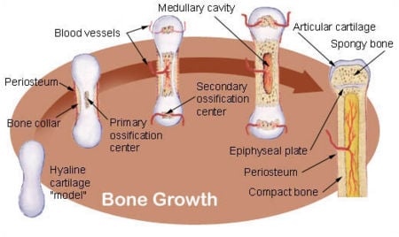
Endochondral ossification occurs in long bones, such as limbs; the bone is formed from cartilage. The steps in endochondral ossification are:
- Development of cartilage model
- Growth of cartilage model
- Development of the primary ossification center
- Development of medullary cavity
- Development of the secondary ossification center
- Formation of articular cartilage and epiphyseal plate
Endochondral ossification begins with points in the cartilage called "primary ossification centers." They mostly appear during fetal development, though a few short bones begin their primary ossification after birth. They are responsible for the formation of the diaphyses of long bones, short bones, and certain parts of irregular bones. Secondary ossification occurs after birth, and forms the epiphyses of long bones and the extremities of irregular and flat bones. The diaphysis and both epiphyses of a long bone are separated by a growing zone of cartilage (the epiphyseal plate). When the child reaches skeletal maturity (18 to 25 years of age), all of the cartilage is replaced by bone, fusing the diaphysis and both epiphyses together (epiphyseal closure).
Bone marrow can be found in almost any bone that holds cancellous tissue. In newborns, all such bones are filled exclusively with red marrow (or hemopoietic marrow), but as the child ages it is mostly replaced by yellow, or "fatty," marrow. In adults, red marrow is mostly found in the flat bones of the skull, the ribs, the vertebrae, and pelvic bones.
"Remodeling" is the process of resorption followed by replacement of bone with little change in shape and occurs throughout a person's life. Its purpose is the release of calcium and the repair of micro-damaged bones (from everyday stress). Repeated stress results in the bone thickening at the points of maximum stress (Wolff's law).
- Bone fracture
- Osteoporosis
- Osteonecrosis
- Osteosarcoma
- Osteogenesis imperfecta
Osteology
The study of bones and teeth is referred to as osteology. It is frequently used in anthropology, archaeology, and forensic science for a variety of tasks. This can include determining the nutrition, health, age, or injury status of the individual the bones were taken from. Preparing fleshed bones for these types of studies can involve maceration—boiling fleshed bones to remove large particles, then hand-cleaning.
Anthropologists and archaeologists also study bone tools made by Homo sapiens and Homo neanderthalensis. Bones can serve a variety of uses, such as projectile points or artistic pigments, and can be made from endoskeletal or external bones such as antler or tusk.
Alternatives to bony endoskeletons
There are several alternatives to mammalary bone seen in nature; though they have some similar functions, they are not completely functionally analogous to bone.
- Exoskeletons offer support, protection, and levers for movement similar to endoskeletal bone. Different types of exoskeletons include shells, carapaces (consisting of calcium compounds or silica) and chitinous exoskelotons.
- A true endoskeleton (that is, protective tissue derived from mesoderm) is also present in echinoderms. Porifera (sponges) possess simple endoskeletons that consist of calcareous or siliceous spicules and a spongin fiber network.
Exposed bone
Bone penetrating the skin and being exposed to the outside can be both a natural process in some animals, and due to injury:
- A deer's antlers are composed of bone
- The extinct predatory fish Dunkleosteus, instead of teeth, had sharp edges of hard exposed bone along its jaws
- A compound fracture occurs when the edges of a broken bone punctures the skin
- Though not strictly exposed, a bird's beak is primarily bone covered in a layer of keratin
Terminology
Several terms are used to refer to features and components of bones throughout the body:
| Bone feature | Definition |
|---|---|
| articular process | A projection that contacts an adjacent bone. |
| articulation | The region where adjacent bones contact each other—a joint. |
| canal | A long, tunnel-like foramen, usually a passage for notable nerves or blood vessels. |
| condyle | A large, rounded articular process. |
| crest | A prominent ridge. |
| eminence | A relatively small projection or bump. |
| epicondyle | A projection near to a condyle but not part of the joint. |
| facet | A small, flattened articular surface. |
| foramen | An opening through a bone. |
| fossa | A broad, shallow depressed area. |
| fovea | A small pit on the head of a bone. |
| labyrinth | A cavity within a bone. |
| line | A long, thin projection, often with a rough surface. Also known as a ridge. |
| malleolus | One of two specific protuberances of bones in the ankle. |
| meatus | A short canal. |
| process | A relatively large projection or prominent bump.(gen.) |
| ramus | An arm-like branch off the body of a bone. |
| sinus | A cavity within a cranial bone. |
| spine | A relatively long, thin projection or bump. |
| suture | Articulation between cranial bones. |
| trochanter | One of two specific tuberosities located on the femur. |
| tubercle | A projection or bump with a roughened surface, generally smaller than a tuberosity. |
| tuberosity | A projection or bump with a roughened surface. |
Several terms are used to refer to specific features of long bones:
| Bone feature | Definition |
|---|---|
| Diaphysis | The long, relatively straight main body of the bone; region of primary ossification. Also known as the shaft. |
| epiphyses | The end regions of the bone; regions of secondary ossification. |
| epiphyseal plate | The thin disc of hyaline cartilage between the diaphysis and epiphyses; disappears by twenty years of age. Also known as the growth plate. |
| head | The proximal articular end of the bone. |
| neck | The region of bone between the head and the shaft. |
References
ISBN links support NWE through referral fees
- Burkhardt, R. 1971. Bone Marrow and Bone Tissue; Color Atlas of Clinical Histopathology. Berlin: Springer-Verlag. ISBN 3540050590.
- Marieb, E. N. 1998. Human Anatomy & Physiology, 4th ed. Menlo Park, California: Benjamin/Cummings Science Publishing. ISBN 080534196X.
- Tortora, G. J. 1989. Principles of Human Anatomy, 5th ed. New York: Harper & Row, Publishers. ISBN 0060466855.
External links
All links retrieved February 8, 2022.
Credits
New World Encyclopedia writers and editors rewrote and completed the Wikipedia article in accordance with New World Encyclopedia standards. This article abides by terms of the Creative Commons CC-by-sa 3.0 License (CC-by-sa), which may be used and disseminated with proper attribution. Credit is due under the terms of this license that can reference both the New World Encyclopedia contributors and the selfless volunteer contributors of the Wikimedia Foundation. To cite this article click here for a list of acceptable citing formats.The history of earlier contributions by wikipedians is accessible to researchers here:
The history of this article since it was imported to New World Encyclopedia:
Note: Some restrictions may apply to use of individual images which are separately licensed.
↧ Download as ZWI file | Last modified: 02/03/2023 22:42:06 | 115 views
☰ Source: https://www.newworldencyclopedia.org/entry/Bones | License: CC BY-SA 3.0
 ZWI signed:


 KSF
KSF