Cryogenic Electron Microscopy
 From Handwiki
  | This article may require copy editing for grammar and sentence structure. (November 2025) (Learn how and when to remove this template message) |

Cryogenic electron microscopy (cryo-EM) is a transmission electron microscopy technique applied to samples cooled to cryogenic temperatures. Developed in the 1970s, advances in detector technology and software allow biomolecular structures to be imaged at near-atomic resolution.[1] The approach has become a popular alternative to X-ray crystallography or NMR spectroscopy in structural biology.[2]
When scanning biological specimens, sample structure is preserved by embedding the specimens in vitreous ice. An aqueous sample solution is applied to a grid-mesh and plunge-frozen in liquid ethane or a mixture of liquid ethane and propane.[3]
The 2017 Nobel Prize in Chemistry was awarded to Jacques Dubochet, Joachim Frank, and Richard Henderson "for developing cryo-electron microscopy for the high-resolution structure determination of biomolecules in solution."[4] Nature Methods also named cryo-EM as the "Method of the Year" in 2015.[5]
History
Early development
In the 1960s, transmission electron microscopy of biological samples was limited because of radiation damage from the high energy electron beams. Scientists hypothesized that examining specimens at low temperatures would reduce beam-induced radiation damage.[6] Both liquid helium (−269 °C or 4 K or −452 °F) and liquid nitrogen (−195.79 °C or 77 K or −320 °F) were considered as cryogens,[7] however high stability was never achieved. In 1980, Erwin Knapek and Jacques Dubochet published comments on beam damage at cryogenic temperatures sharing observations that:
Thin crystals mounted on carbon film were found to be from 30 to 300 times more beam-resistant at 4 K than at room temperature... Most of our results can be explained by assuming that cryoprotection in the region of 4 K is strongly dependent on the temperature.[8]
However, these results were not reproduced and just two years later amendments were published,[9] along with a commentary in Nature,[10] indicating that the beam resistance was less significant than anticipated. The protection gained at 4 K was closer to "tenfold for standard samples of L-valine", than what was previously stated.[10] While cryo-EM samples are routinely collected at liquid nitrogen temperatures,[11] work has continued to understand sample behavior at liquid helium temperatures.[12][13][11]
In 1981 scientists at the European Molecular Biology Laboratory, reported the first successful cryo-EM.[14] Researchers used a thin film of vitrified pure water a hydrophilic carbon film that was rapidly plunged into cryogen (liquid propane or liquid ethane cooled to 77 K). The thin layer of amorphous ice was less than 1 μm thick and an electron diffraction pattern confirmed the presence of amorphous/vitreous ice. In 1984 the group demonstrated the power of cryo-EM in structural biology by analysing vitrified adenovirus type 2, T4 bacteriophage, Semliki Forest virus, Bacteriophage CbK, and Vesicular-Stomatitis-Virus.[15] The paper marked the origin of Cryo-EM, and the technique has become routine in laboratories throughout the world.
The energy of the electrons used for imaging (80–300 kV) can break covalent bonds in organic and biological samples.[16] Imaging biological specimens requires minimising electron exposure. Low exposures require images of thousands or millions of frozen molecules be selected, aligned, and averaged to obtain high-resolution maps, using specialized software. The 2012 introduction of direct electron detectors and better computational algorithms significantly improved structural features.[17]
Recent advancements
Direct Electron Detectors, and more powerful imaging algorithms allow macromolecular structures to be determined at near-atomic resolution.[18] Imaged macromolecules include viruses, ribosomes, mitochondria, ion channels, and enzyme complexes. Starting in 2018, cryo-EM could be applied to structures as small as hemoglobin (64 kDa)[19] with resolutions up to 1.8 Å.[20] In 2019, cryo-EM structures grew to 2.5% of structures deposited in the Protein Data Bank.[21][22] Cryo-EM can be used for cryo-electron tomography (cryo-ET), creating 3D reconstructions of samples from tilted 2D images.
The 2010s saw drastic advancements of electron cameras, including to direct electron detectors, causing a "resolution revolution"[23] pushing the resolution barrier beneath the crucial ~2-3 Å limit to resolve amino acid position and orientation.[24]
Richard Henderson (MRC Laboratory of Molecular Biology, Cambridge, UK) formed a consortium with engineers at the Rutherford Appleton Laboratory and scientists at the Max Planck Society to fund and develop a first prototype. The consortium then joined forces with the electron microscope manufacturer FEI to roll out and market the new design. At about the same time, Gatan Inc. of Pleasanton, California came out with a similar detector designed by Peter Denes (Lawrence Berkeley National Laboratory) and David Agard (University of California, San Francisco). A third type of camera was developed by Nguyen-Huu Xuong at the Direct Electron company (San Diego, California).[23]
Recent advancements in protein-based imaging scaffolds assist with sample orientation bias and size limit. Though the minimum size for Cryo-EM remains undetermined, proteins smaller than ~50 kDa generally have too low a signal-to-noise ratio (SNR) to resolve protein particles, making 3D reconstruction difficult or impossible.[25][26] Multiple techniques have been reported to improve SNR when determining the structures of small proteins.[27][28] Based on high-affinity DARPins, nanobodies, antibody fragments,[29] these methods rigidly bind the target protein and thereby increase the effective particle size and introduce symmetry to improve SNR for Cryo-EM map reconstruction. An advantage of Cryo-EM over crystallization is that it requires much less sample material. This makes it easier to determine structures of proteins that cannot be isolated with high yield.
2017 Nobel Prize in Chemistry
In recognition of the impact cryo-EM has had on biochemistry, three scientists, Jacques Dubochet, Joachim Frank and Richard Henderson, were awarded the Nobel Prize in Chemistry "for developing cryo-electron microscopy for the high-resolution structure determination of biomolecules in solution."[4]
Techniques
Mode of electron microscopy
Cryogenic transmission electron microscopy
Cryogenic transmission electron microscopy (cryo-TEM) is a transmission electron microscopy technique that is used in structural biology and materials science. Colloquially, the term "cryogenic electron microscopy" or its shortening "cryo-EM" refers to cryogenic transmission electron microscopy by default, as the vast majority of cryo-EM is done in transmission electron microscopes, rather than scanning electron microscopes.
Correlative light cryo-TEM and cryo-ET
In 2019, correlative light cryo-TEM and cryo-ET were used to observe tunnelling nanotubes (TNTs) in neuronal cells.[30]
Scanning electron cryomicroscopy
Scanning electron cryomicroscopy (cryoSEM) is a scanning electron microscopy technique with a scanning electron microscope's cold stage in a cryogenic chamber.
Technique of use and data analysis
Electron cryotomography
In electron cryotomography (cyro-ET), many pictures of a sample are taken from different angles using a tilting mechanism. The images are combined to create a 3D model (map) of ~1–4 nm resolution.[31]
Single particle analysis

SPA or single-particle cyro-EM is the method used to obtain near-atomic resolution (<1 nm) models of biomolecules. It is what the 2017 Nobel Prize refers to. In SPA, a large collection of cyro-TEM images are automatically sorted into classes. Within each class, the images are combined to reduce noise and to create a 3D model of the class of particles, a 3D "map". The main innovation compared to cyro-ET is the combination of images from similar objects.[32]
When combined with a knowledge of time progression, the result is time-resolved cyro-TEM.[33][34][35]
Comparisons to X-ray crystallography
Traditionally, X-ray crystallography has been the most popular technique for determining the 3D structures of biological molecules.[36] However, the aforementioned improvements in cryo-EM have increased its popularity as a tool for examining the details of biological molecules. Since 2010, yearly cryo-EM structure deposits have outpaced X-ray crystallography.[37] Though X-ray crystallography has drastically more total deposits due to a decades-longer history, total deposits of the two methods are projected to eclipse around 2035.[37]
The resolution of X-ray crystallography is limited by crystal homogeneity,[38] and coaxing biological molecules with unknown ideal crystallization conditions into a crystalline state can be very time-consuming, in extreme cases taking months or even years.[39] To contrast, sample preparation in cryo-EM may require several rounds of screening and optimization to overcome issues such as protein aggregation and preferred orientations,[40][41] but it does not require the sample to form a crystal, rather samples for cryo-EM are flash-frozen and examined in their near-native states.[42]
According to Proteopedia, the median resolution achieved by X-ray crystallography (as of May 19, 2019) on the Protein Data Bank is 2.05 Å,[38] and the highest resolution achieved on record (as of September 30, 2022) is 0.48 Å.[43] As of 2020, the majority of the protein structures determined by cryo-EM (single particle analysis) are at a lower resolution of 3–4 Å.[44] However, as of 2020, the best cryo-EM resolution has been recorded at 1.22 Å,[41] making it a competitor in resolution in some cases.
Electron crystallography
Similar to X-ray crystallography used to determine the crystal structure of molecules of different sizes (from small molecules to large biomolecular complexes) using the X-ray diffraction pattern, electrons can also produce a electron diffraction pattern from a crystal. Work in this area has a long history dating back to early work such as the determination of the positions of hydrogen atoms in NH4Cl crystals by W. E. Laschkarew and I. D. Usykin in 1933,[45]
Cyro-EC is typically done with 3D crystals, but it has also been used in analysis of two-dimensional crystals and analysis of helical filaments or tubes.
Microcrystal electron diffraction (MicroED) is a version of electron crystallography that works with crystals a billion times smaller than what X-ray diffraction requires. It has been used to determine the structure of large biomolecules (proteins, nucleic acids, their complexes).[46] It is also very useful in studying small molecules, from peptides to simpler compounds.[47]
Specimen handling for imaging
(This section does not apply to electron crystallography.)
Biological specimens
Thin film
The biological material is spread on an electron microscopy grid and is preserved in a frozen-hydrated state by rapid freezing, usually in liquid ethane near liquid nitrogen temperature. By maintaining specimens at liquid nitrogen temperature or colder, they can be introduced into the high-vacuum of the electron microscope column. Most biological specimens are extremely radiosensitive, so they must be imaged with low-dose techniques (usefully, the low temperature of transmission electron cryomicroscopy provides an additional protective factor against radiation damage).
Consequently, the images are extremely noisy. For some biological systems it is possible to average images to increase the signal-to-noise ratio and retrieve high-resolution information about the specimen using the technique known as single particle analysis. This approach in general requires that the things being averaged are identical, although some limited conformational heterogeneity can now be studied (e.g. ribosome). Three-dimensional reconstructions from CryoTEM images of protein complexes and viruses have been solved to sub-nanometer or near-atomic resolution, allowing new insights into the structure and biology of these large assemblies.
Analysis of ordered arrays of protein, such as 2-D crystals of transmembrane proteins or helical arrays of proteins, also allows a kind of averaging which can provide high-resolution information about the specimen. This technique is called electron crystallography.
Vitreous sections
The thin film method is limited to thin specimens (typically < 500 nm) because the electrons cannot cross thicker samples without multiple scattering events. Thicker specimens can be vitrified by plunge freezing (cryofixation) in ethane (up to tens of μm in thickness) or more commonly by high pressure freezing (up to hundreds of μm). They can then be cut in thin sections (40 to 200 nm thick) with a diamond knife in a cryoultramicrotome at temperatures lower than −135 °C (devitrification temperature). The sections are collected on an electron microscope grid and are imaged in the same manner as specimen vitrified in thin film. This technique is called transmission electron cryomicroscopy of vitreous sections (CEMOVIS) or transmission electron cryomicroscopy of frozen-hydrated sections.
Material specimens
In addition to allowing vitrified biological samples to be imaged, CryoTEM can also be used to image material specimens that are too volatile in vacuum to image using standard, room temperature electron microscopy. For example, vitrified sections of liquid-solid interfaces can be extracted for analysis by CryoTEM,[48] and sulfur, which is prone to sublimation in the vacuum of electron microscopes, can be stabilized and imaged in CryoTEM.[49]
Image processing in cryo-TEM
Even though in the majority of approaches in electron microscopy one tries to get the best resolution image of the material, it is not always the case in cryo-TEM. Besides all the benefits of high resolution images, the signal to noise ratio remains the main hurdle that prevents assigning orientation to each particle. For example, in macromolecule complexes, there are several different structures that are being projected from 3D to 2D during imaging and if they are not distinguished the result of image processing will be a blur. That is why the probabilistic approaches become more powerful in this type of investigation.[50] There are two popular approaches that are widely used nowadays in cryo-EM image processing, the maximum likelihood approach that was discovered in 1998[51] and relatively recently adapted Bayesian approach.[52]
The maximum likelihood estimation approach comes to this field from the statistics. Here, all the possible orientations of particles are summed up to get the resulting probability distribution. We can compare this to a typical least square estimation where particles get exact orientations per image.[53] This way, the particles in the sample get "fuzzy" orientations after calculations, weighted by corresponding probabilities. The whole process is iterative and with each next iteration the model gets better. The good conditions for making the model that closely represent the real structure is when the data does not have too much noise and the particles do not have any preferential direction. The main downside of maximum likelihood approach is that the result depends on the initial guess and model optimization can sometimes get stuck at local minimum.[54]
The Bayesian approach that is now being used in cryo-TEM is empirical by nature. This means that the distribution of particles is based on the original dataset. Similarly, in the usual Bayesian method there is a fixed prior probability that is changed after the data is observed. The main difference from the maximum likelihood estimation lies in special reconstruction term that helps smoothing the resulting maps while also decreasing the noise during reconstruction.[53] The smoothing of the maps occurs through assuming prior probability to be a Gaussian distribution and analyzing the data in the Fourier space. Since the connection between the prior knowledge and the dataset is established, there is less chance for human factor errors which potentially increases the objectivity of image reconstruction.[52]
With emerging new methods of cryo-TEM imaging and image reconstruction the new software solutions appear that help to automate the process. After the empirical Bayesian approach have been implemented in the open source computer program RELION (REgularized LIkelihood OptimizatioN) for 3D reconstruction,[55][56] the program became widespread in the cryo-TEM field. It offers a range of corrections that improve the resolution of reconstructed images, allows implementing versatile scripts using python language and executes the usual tasks of 2D/3D model classifications or creating de novo models.[57][58]
Gallery
-
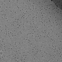
-
 Structure of alcohol oxidase from Pichia pastoris by Cryo-EM
 -
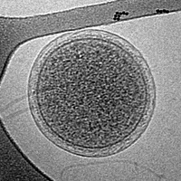 Cryo-EM image of an intact ARMAN cell from an Iron Mountain biofilm. Image width is 576 nm.
 -
![Cryo-EM image of the CroV giant marine virus (scale bar represents 200 nm)[59]](tmp/UlaNASqfesKh/data/media/images/200px-CroV_TEM_(cropped).jpg) Cryo-EM image of the CroV giant marine virus
Cryo-EM image of the CroV giant marine virus
(scale bar represents 200 nm)[59]


![Cryo-EM image of the CroV giant marine virus (scale bar represents 200 nm)[59]](https://handwiki.org/wiki/File:CroV_TEM_(cropped).jpg)
See also
 |
Wikibooks has a book on the topic of: Software Tools For Molecular Microscopy |
- Cryogenic scanning electron microscopy
- EM Data Bank
- Resolution (electron density)
- Single particle analysis
- Cryofixation
- Cryo bio-crystallography
- Electron tomography (ET)
- Virus crystallisation
References
- ↑ "A primer to single-particle cryo-electron microscopy". Cell 161 (3): 438–449. April 2015. doi:10.1016/j.cell.2015.03.050. PMID 25910204.
- ↑ "Structural biology: How proteins got their close-up". Knowable Magazine. 1 March 2022. doi:10.1146/knowable-022822-1. https://knowablemagazine.org/article/living-world/2022/structural-biology-how-proteins-got-their-closeup. Retrieved 25 March 2022.
- ↑ "An improved cryogen for plunge freezing". Microscopy and Microanalysis 14 (5): 375–379. October 2008. doi:10.1017/S1431927608080781. PMID 18793481. Bibcode: 2008MiMic..14..375T.
- ↑ 4.0 4.1 "The Nobel Prize in Chemistry 2017" (in en-US). https://www.nobelprize.org/prizes/chemistry/2017/press-release/.
- ↑ "Cryo-electron tomography". Nature Methods 14 (1): 34. January 2017. doi:10.1038/nmeth.4115. ISSN 1548-7091.
- ↑ "Ups and downs in early electron cryo-microscopy". PLOS Biology 16 (4). April 2018. doi:10.1371/journal.pbio.2005550. PMID 29672565.
- ↑ "Liquid helium cooled tilting stage for an electron microscope". Review of Scientific Instruments 34 (5): 582–583. 1963.
- ↑ "Beam damage to organic material is considerably reduced in cryo-electron microscopy". Journal of Molecular Biology 141 (2): 147–161. August 1980. doi:10.1016/0022-2836(80)90382-4. PMID 7441748.
- ↑ Lepault, J.; Dubochet, J.; Dietrich, I.; Knapek, E.; Zeitler, E. (January 1983). "Amendment to: Electron beam damage to organic specimens at liquid helium temperature". Journal of Molecular Biology 163 (3): 511. doi:10.1016/0022-2836(83)90073-6.
- ↑ 10.0 10.1 "Cryo-transmission microscopy Fading hopes". Nature 299 (5882): 386–387. 30 September 1982. doi:10.1038/299386c0. Bibcode: 1982Natur.299..386N.
- ↑ 11.0 11.1 Dickerson, JL; Naydenova, K; Peet, MJ; Wilson, H; Nandy, B; McMullan, G; Morrison, R; Russo, CJ (29 April 2025). "Reducing the effects of radiation damage in cryo-EM using liquid helium temperatures.". Proceedings of the National Academy of Sciences of the United States of America 122 (17). doi:10.1073/pnas.2421538122. PMID 40261934.
- ↑ Pfeil-Gardiner, Olivia; Mills, Deryck J.; Vonck, Janet; Kuehlbrandt, Werner (1 November 2019). "A comparative study of single-particle cryo-EM with liquid-nitrogen and liquid-helium cooling". IUCrJ 6 (6): 1099–1105. doi:10.1107/S2052252519011503.
- ↑ Dickerson, Joshua L; Russo, Christopher J (25 July 2025). "A Physical Theory For Cryo-EM At Liquid-Helium Temperatures". Microscopy and Microanalysis 31 (Supplement_1). doi:10.1093/mam/ozaf048.514.
- ↑ "Vitrification of Pure Water for Electron Microscopy" (in en). Journal of Microscopy 124 (3): 3–4. December 1981. doi:10.1111/j.1365-2818.1981.tb02483.x.
- ↑ "Cryo-electron microscopy of viruses". Nature 308 (5954): 32–36. March 1984. doi:10.1038/308032a0. PMID 6322001. Bibcode: 1984Natur.308...32A. https://serval.unil.ch/notice/serval:BIB_BEC796503260.
- ↑ Garman, Elspeth F.; Owen, Robin Leslie (2006-01-01). "Cryocooling and radiation damage in macromolecular crystallography". Acta Crystallographica Section D Biological Crystallography 62 (1): 32–47. doi:10.1107/S0907444905034207. ISSN 0907-4449. https://journals.iucr.org/paper?S0907444905034207.
- ↑ "The revolution will not be crystallized: a new method sweeps through structural biology". Nature 525 (7568): 172–4. September 2015. doi:10.1038/525172a. PMID 26354465. Bibcode: 2015Natur.525..172C.
- ↑ "Cryo-electron microscopy for structural analysis of dynamic biological macromolecules". Biochimica et Biophysica Acta (BBA) - General Subjects 1862 (2): 324–334. Feb 2018. doi:10.1016/j.bbagen.2017.07.020. PMID 28756276.
- ↑ "Cryo-EM structure of haemoglobin at 3.2 Å determined with the Volta phase plate". Nature Communications 8. June 2017. doi:10.1038/ncomms16099. PMID 28665412. Bibcode: 2017NatCo...816099K.
- ↑ "Breaking Cryo-EM Resolution Barriers to Facilitate Drug Discovery". Cell 165 (7): 1698–1707. June 2016. doi:10.1016/j.cell.2016.05.040. PMID 27238019.
- ↑ "PDB Data Distribution by Experimental Method and Molecular Type". https://www.rcsb.org/stats/summary.
- ↑ "PDB Statistics: Growth of Structures from 3DEM Experiments Released per Year". https://www.rcsb.org/stats/growth/em.
- ↑ 23.0 23.1 Kühlbrandt, Werner (2014-03-28). "The Resolution Revolution" (in en). Science 343 (6178): 1443–1444. doi:10.1126/science.1251652. ISSN 0036-8075. PMID 24675944. Bibcode: 2014Sci...343.1443K. https://www.science.org/doi/10.1126/science.1251652.
- ↑ Kuster, Daniel J.; Liu, Chengyu; Fang, Zheng; Ponder, Jay W.; Marshall, Garland R. (2015-04-20). "High-Resolution Crystal Structures of Protein Helices Reconciled with Three-Centered Hydrogen Bonds and Multipole Electrostatics" (in en). PLOS ONE 10 (4). doi:10.1371/journal.pone.0123146. ISSN 1932-6203. PMID 25894612. Bibcode: 2015PLoSO..1023146K.
- ↑ Henderson, Richard (May 1995). "The potential and limitations of neutrons, electrons and X-rays for atomic resolution microscopy of unstained biological molecules". Quarterly Reviews of Biophysics 28 (2): 171–193. doi:10.1017/s003358350000305x.
- ↑ Herzik, Mark A.; Wu, Mengyu; Lander, Gabriel C. (2019-03-04). "High-resolution structure determination of sub-100 kDa complexes using conventional cryo-EM" (in en). Nature Communications 10 (1): 1032. doi:10.1038/s41467-019-08991-8. ISSN 2041-1723. PMID 30833564. Bibcode: 2019NatCo..10.1032H.
- ↑ Wentinck, Koen; Gogou, Christos; Meijer, Dimphna H. (2022). "Putting on molecular weight: Enabling cryo-EM structure determination of sub-100-kDa proteins". Current Research in Structural Biology 4: 332–337. doi:10.1016/j.crstbi.2022.09.005.
- ↑ Gilman, Morgan S. A.; Skiba, Meredith A. (15 August 2025). "Hidden gems bring proteins into view". Nature Chemical Biology. doi:10.1038/s41589-025-01959-4.
- ↑
- Wu, Shenping; Avila-Sakar, Agustin; Kim, JungMin; Booth, David S.; Greenberg, Charles H.; Rossi, Andrea; Liao, Maofu; Li, Xueming et al. (April 2012). "Fabs Enable Single Particle cryoEM Studies of Small Proteins". Structure 20 (4): 582–592. doi:10.1016/j.str.2012.02.017.Mukherjee, Somnath; Erramilli, Satchal K.; Ammirati, Mark; Alvarez, Frances J. D.; Fennell, Kimberly F.; Purdy, Michael D.; Skrobek, Blazej M.; Radziwon, Katarzyna et al. (27 March 2020). "Synthetic antibodies against BRIL as universal fiducial marks for single−particle cryoEM structure determination of membrane proteins". Nature Communications 11 (1). doi:10.1038/s41467-020-15363-0.Xie, Pujun; Li, Yan; Lamon, Gaëlle; Kuang, Huihui; Wang, Da-Neng; Traaseth, Nathaniel J. (2 January 2025). "A fiducial-assisted strategy compatible with resolving small MFS transporter structures in multiple conformations using cryo-EM". Nature Communications 16 (1). doi:10.1038/s41467-024-54986-5."Cryo-EM structure determination of small therapeutic protein targets at 3 Å-resolution using a rigid imaging scaffold". Proceedings of the National Academy of Sciences of the United States of America 120 (37). September 2023. doi:10.1073/pnas.2305494120. PMID 37669364. Bibcode: 2023PNAS..12005494C.Ackle, Fabian; Thavarasah, Sujani; Earp, Jennifer C.; Seeger, Markus A. (19 March 2025). "Rigid enlargement of sybodies with antibody fragments for cryo-EM analyses of small membrane proteins". Scientific Reports 15 (1). doi:10.1038/s41598-025-92950-5.Wu, Xudong; Rapoport, Tom A. (12 October 2021). "Cryo-EM structure determination of small proteins by nanobody-binding scaffolds (Legobodies)". Proceedings of the National Academy of Sciences 118 (41). doi:10.1073/pnas.2115001118.Yi, Gangshun; Mamalis, Dimitrios; Ye, Mingda; Carrique, Loic; Fairhead, Michael; Li, Huanyu; Duerr, Katharina L.; Zhang, Peijun et al. (15 August 2025). "Covalently constrained 'Di-Gembodies' enable parallel structure solutions by cryo-EM". Nature Chemical Biology. doi:10.1038/s41589-025-01972-7.Kung, Jennifer E.; Johnson, Matthew C.; Tegunov, Dimitry; Jao, Christine C.; Wu, Ping; Oh, Angela; Lin, May; Daria, Jose M. et al. (30 September 2025). "Disulfide-constrained Fabs overcome target size limitation for high-resolution single particle cryoEM". Nature Communications 16 (1). doi:10.1038/s41467-025-63766-8.
- ↑ "Correlative cryo-electron microscopy reveals the structure of TNTs in neuronal cells". Nature Communications 10 (1): 342. January 2019. doi:10.1038/s41467-018-08178-7. PMID 30664666. Bibcode: 2019NatCo..10..342S.
- ↑ Bäuerlein, Felix J. B.; Baumeister, Wolfgang (2021-10-01). "Towards Visual Proteomics at High Resolution". Journal of Molecular Biology. From Protein Sequence to Structure at Warp Speed: How Alphafold Impacts Biology 433 (20). doi:10.1016/j.jmb.2021.167187. ISSN 0022-2836. PMID 34384780.
- ↑ "Single-particle cryo-EM-How did it get here and where will it go". Science 361 (6405): 876–880. August 2018. doi:10.1126/science.aat4346. PMID 30166484. Bibcode: 2018Sci...361..876C.
- ↑ "Key Intermediates in Ribosome Recycling Visualized by Time-Resolved Cryoelectron Microscopy". Structure 24 (12): 2092–2101. December 2016. doi:10.1016/j.str.2016.09.014. PMID 27818103.
- ↑ "A Fast and Effective Microfluidic Spraying-Plunging Method for High-Resolution Single-Particle Cryo-EM". Structure 25 (4): 663–670.e3. April 2017. doi:10.1016/j.str.2017.02.005. PMID 28286002.
- ↑ "Structural dynamics of ribosome subunit association studied by mixing-spraying time-resolved cryogenic electron microscopy". Structure 23 (6): 1097–105. June 2015. doi:10.1016/j.str.2015.04.007. PMID 26004440.
- ↑ "x ray crystallography". Molecular Pathology 53 (1): 8–14. February 2000. doi:10.1136/mp.53.1.8. PMID 10884915.
- ↑ 37.0 37.1 Chiu, Wah; Schmid, Michael F.; Pintilie, Grigore D.; Lawson, Catherine L. (January 2021). "Evolution of standardization and dissemination of cryo-EM structures and data jointly by the community, PDB, and EMDB". Journal of Biological Chemistry 296. doi:10.1016/j.jbc.2021.100560. ISSN 0021-9258. PMID 33744287.
- ↑ 38.0 38.1 "Resolution - Proteopedia, life in 3D". https://proteopedia.org/wiki/index.php/Resolution#Confusion_of_high_vs._low_resolution.
- ↑ "Revolutionary cryo-EM is taking over structural biology". Nature 578 (7794): 201. February 2020. doi:10.1038/d41586-020-00341-9. PMID 32047310. Bibcode: 2020Natur.578..201C.
- ↑ Lyumkis, Dmitry (2019-03-29). "Challenges and opportunities in cryo-EM single-particle analysis". Journal of Biological Chemistry 294 (13): 5181–5197. doi:10.1074/jbc.rev118.005602. ISSN 0021-9258. PMID 30804214.
- ↑ 41.0 41.1 "Single-particle cryo-EM at atomic resolution". Nature 587 (7832): 152–156. November 2020. doi:10.1038/s41586-020-2829-0. PMID 33087931. Bibcode: 2020Natur.587..152N.
- ↑ "How cryo-electron microscopy and X-ray crystallography complement each other". Protein Science 26 (1): 32–39. January 2017. doi:10.1002/pro.3022. PMID 27543495.
- ↑ "Crystal structure of small protein crambin at 0.48 Å resolution". Acta Crystallographica. Section F, Structural Biology and Crystallization Communications 67 (Pt 4): 424–428. April 2011. doi:10.1107/S1744309110052607. PMID 21505232.
- ↑ "Atomic-resolution protein structure determination by cryo-EM". Nature 587 (7832): 157–161. November 2020. doi:10.1038/s41586-020-2833-4. PMID 33087927. Bibcode: 2020Natur.587..157Y.
- ↑ Laschkarew, W. E.; Usyskin, I. D. (1933). "Die Bestimmung der Lage der Wasserstoffionen im NH4Cl-Kristallgitter durch Elektronenbeugung" (in de). Zeitschrift für Physik 85 (9–10): 618–630. doi:10.1007/BF01331003. ISSN 1434-6001. Bibcode: 1933ZPhy...85..618L. http://link.springer.com/10.1007/BF01331003.
- ↑ "The collection of MicroED data for macromolecular crystallography". Nature Protocols 11 (5): 895–904. May 2016. doi:10.1038/nprot.2016.046. PMID 27077331.
- ↑ "The CryoEM Method MicroED as a Powerful Tool for Small Molecule Structure Determination". ACS Central Science 4 (11): 1587–1592. November 2018. doi:10.1021/acscentsci.8b00760. PMID 30555912.
- ↑ "Site-Specific Preparation of Intact Solid-Liquid Interfaces by Label-Free In Situ Localization and Cryo-Focused Ion Beam Lift-Out". Microscopy and Microanalysis 22 (6): 1338–1349. December 2016. doi:10.1017/S1431927616011892. PMID 27869059. Bibcode: 2016MiMic..22.1338Z.
- ↑ "Characterization of Sulfur and Nanostructured Sulfur Battery Cathodes in Electron Microscopy Without Sublimation Artifacts". Microscopy and Microanalysis 23 (1): 155–162. February 2017. doi:10.1017/S1431927617000058. PMID 28228169. Bibcode: 2017MiMic..23..155L. https://zenodo.org/record/889883.
- ↑ Cheng, Yifan (2018-08-31). "Single-particle cryo-EM—How did it get here and where will it go" (in en). Science 361 (6405): 876–880. doi:10.1126/science.aat4346. ISSN 0036-8075. PMID 30166484. Bibcode: 2018Sci...361..876C.
- ↑ Sigworth, F.J. (1998). "A Maximum-Likelihood Approach to Single-Particle Image Refinement" (in en). Journal of Structural Biology 122 (3): 328–339. doi:10.1006/jsbi.1998.4014. PMID 9774537.
- ↑ 52.0 52.1 Scheres, Sjors H.W. (January 2012). "A Bayesian View on Cryo-EM Structure Determination" (in en). Journal of Molecular Biology 415 (2): 406–418. doi:10.1016/j.jmb.2011.11.010. PMID 22100448.
- ↑ 53.0 53.1 Nogales, Eva; Scheres, Sjors H.W. (May 2015). "Cryo-EM: A Unique Tool for the Visualization of Macromolecular Complexity". Molecular Cell 58 (4): 677–689. doi:10.1016/j.molcel.2015.02.019. ISSN 1097-2765. PMID 26000851.
- ↑ Sigworth, Fred J. (2016-02-01). "Principles of cryo-EM single-particle image processing". Microscopy 65 (1): 57–67. doi:10.1093/jmicro/dfv370. ISSN 2050-5698. PMID 26705325.
- ↑ Scheres, Sjors H. W. (2012-12-01). "RELION: Implementation of a Bayesian approach to cryo-EM structure determination" (in en). Journal of Structural Biology 180 (3): 519–530. doi:10.1016/j.jsb.2012.09.006. ISSN 1047-8477. PMID 23000701.
- ↑ "RELION: Image-processing software for cryo-electron microscopy". 3dem. 27 October 2023. https://github.com/3dem/relion.
- ↑ Bai, Xiao-chen; McMullan, Greg; Scheres, Sjors H.W (January 2015). "How cryo-EM is revolutionizing structural biology". Trends in Biochemical Sciences 40 (1): 49–57. doi:10.1016/j.tibs.2014.10.005. ISSN 0968-0004. PMID 25544475.
- ↑ Zivanov, Jasenko; Nakane, Takanori; Forsberg, Björn O; Kimanius, Dari; Hagen, Wim JH; Lindahl, Erik; Scheres, Sjors HW (2018-11-09). Egelman, Edward H; Kuriyan, John. eds. "New tools for automated high-resolution cryo-EM structure determination in RELION-3". eLife 7. doi:10.7554/eLife.42166. ISSN 2050-084X. PMID 30412051.
- ↑ Xiao, C., Fischer, M.G., Bolotaulo, D.M., Ulloa-Rondeau, N., Avila, G.A., and Suttle, C.A. (2017) "Cryo-EM reconstruction of the Cafeteria roenbergensis virus capsid suggests novel assembly pathway for giant viruses". Scientific Reports, 7: 5484. doi:10.1038/s41598-017-05824-w.
| Basics | |||||||
|---|---|---|---|---|---|---|---|
| Electron interaction with matter | |||||||
| Instrumentation | |||||||
| Microscopes |
| ||||||
| Techniques |
| ||||||
| Others |
| ||||||


 |  |
Categories: [Cell biology] [Protein structure] [Scientific techniques]
↧ Download as ZWI file | Last modified: 08/23/2026 02:29:13 | 3 views
☰ Source: https://handwiki.org/wiki/Engineering:Cryogenic_electron_microscopy | License: CC BY-SA 3.0
 KSF
KSF