Central Nervous System
 From Conservapedia
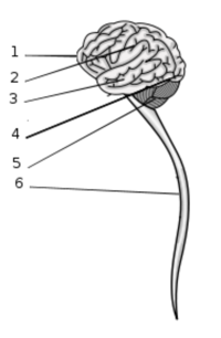
1. Frontal lobe
2. Parietal lobe
3. Temporal lobe
4. Occipital lobe
5. Cerebellum
6. Spinal cord
The central nervous system (CNS) in its most simple divisions consists of all nervous tissue in the spinal cord and the brain. It is the largest division of the nervous system and has primary control over all organism behavior. Much of our understanding of the central nervous system stems from the computational theory of mind. In modern cognitive psychology and neuroscience the CNS is viewed as an information processing system similar to a modern computer. It is theorized that all thought, conscious and unconsciousness, as well as all behavior is controlled by the same underlying hardware of neurons. The CNS is also viewed as being inherently modular with specific functions being localized to specific areas.
Contents
Structure[edit]
The central nervous system is divided into seven major divisions. The first is the spinal cord which integrates with the brain stem and runs down the center of the vertebrate column. The brain itself is composed of six regions the medulla, pons, cerebellum, midbrain, diencephalon, and the cerebral hemispheres (sometimes referred to as telencephalon). Each of these regions can be further subdivided into many anatomically and functionally distinct areas.
The central nervous system is covered by three tissue layers called the meninges. The three meninges tissues are the dura mater, arachnoid mater, and pia mater. The dura mater is the thickest and toughest tissue and the outermost layer. It is primarily thought to serve as a protection layer for the underlying nervous tissue. The arachnoid mater is connected loosely to the dura mater. There is a space between the dura mater and the arachnoid mater called the subdural space. The pia mater is a very thin and delicate layer of tissue that adheres to the surface of the nervous tissue. The space between the archnoid mater and the pia mater is called the subarachnoid space. Arachnoid mater filaments pass through this space giving a web like appearance to the area (hence the term arachnoid).
Beyond protection the meninges tissue is also important for the circulatory health of the central nervous system. The veins and arteries that serve the CNS are located in the subarachnoid space. There are also large, low-pressure blood vessels that serve as the return path for cerebral venous blood located in the dura mater.
The CNS contains several cavities called the ventrical system. Tissue surrounding the ventrical system is the primary producer of cerebrospinal fluid which surrounds the CNS providing a medium for communication between neurons and as a cushion in case of trauma.
Neurons and axons are distributed non-uniformly through the CNS. The cell body and dendrites of the neurons cluster in cortical regions which are essentially flattened sheets of cells called laminae. These areas are located primarily on the cerebral hemispheres or in areas beneath the surface of all the sub-divisions of the CNS called nuclei. Axons are then distributed through out the system in regions called tracts. In live tissue the regions populated by cell bodies and dendrites appear gray while the tract's of axons appear white due to myelin sheaths. These areas are called gray matter and white matter respectively.
Spinal cord[edit]
The spinal cord is the primary tract for sensory information coming in from the periphery to the brain and voluntary muscle control signals moving from the brain to the periphery. Information traveling to the brain moves through a series of axons called the afferent nerves and information traveling to the periphery move through axons called the efferent nerves. The spinal cord can also help processes and integrate sensory information before passing it on. In a few cases it can receive input and coordinate behavioral responses without ever recruiting the brain, these behaviors are commonly called reflexes.
The spinal cord is segmental with the overall organization being modular, all the segments are the same basic structure repeated in tandem. Each segment has a pair of nerves, the dorsal root and the ventral root. The dorsal root contains only the sensory, afferent axons. The ventral root contains the motor, or efferent axons.
The brain stem and cerebellum[edit]
The medulla, pons and midbrain comprise what is commonly called the brain stem. The primary functions assigned to the brain stem include receiving sensory information from cranial structures and controlling the muscles of the head. The brain stem also is the major conduit for information coming into and out of the brain. Finally, the brain stem is the primary integrator and controller of many arousal and basic functions. The main function of the cerebellum appears to be to regulate eye and limb movements. Also it maintains posture and balance.
The diencephalon[edit]
The diencephalon is divided into the thalamus and hypothalamus. The thalamus serves to integrate sensory information and pass it on to the cerebral cortex. The hypothalamus receives and integrates information from the autonomic system and controls the endocrine system as well.
The cerebral hemisphere[edit]
The cerebral hemisphere is a highly developed and complex structure. Its divided into four major sections: cerebral cortex, hippocampal formation, amygdala, and the basal ganglia. Most behavior that is viewed as uniquely human is controlled by these four structures.
The hippocampal formation is primarily used in memory and learning, the amygdala controls emotion and the organisms response to stress. Together with some parts of the brain stem these structures make up the limbic system which plays a very large role in mood and psychological dysfunctions. The basal ganglia is important in movement and muscle control, it is the area of the brain that is damaged with Parkinson's Disease. It is also important in cognition and emotion and seems to be a key area involved in addiction.
The cerebral cortex is located on the surface of the brain and has a highly complex three dimensional structure of convoluted nervous tissue. It is essentially a long sheet that has been folded up again and again. These folds are thought to be an adaptation to squeezing in large amounts of nervous tissue in the limited space of the cranial skull. These folds create a wave pattern with the elevated portion of the tissue being called gyri and the separations between called sulci. Some sulci are particularly deep and called fissures. The two hemispheres of the brain are separated by a large fissure called the sagittal fissure. The hemispheres are connected by a structure called the corpus callosum.
The cerebral cortex is divided into four lobes, the frontal lobe, the temporal lobe, the parietal lobe, and the occipital lobe. The names for these lobes correspond to the cranial bones that overlie them. The frontal lobe is primarily involved in planning, speech, cognition and emotions. The parietal lobe is involved in perceptions of touch, pain and limb position. The occipital lobe has almost the singular function of decoding visual information and is the site of the primary visual cortex. The temporal lobe controls memory, emotion and various sensory functions.
Development[edit]
Vertebrate embryos form three primary layers of tissue. The ectoderm (outermost layer), mesoderm (middle layer) and endoderm (inner most layer). The nervous tissue is formed from a region of the ectoderm called the neural plate. The formation of the nervous system is triggered by signaling molecules that come from the mesoderm. This process is called neural induction.
The neural plate runs along the midline of the embryo. Cell division is disproportionately prolific along the midline which forms a grove called the neural grove. This grove deepens and eventually closes forming a hollow tube called the neural tube. Early in development portions at the top of the neural tube swell up forming three hollow vesicles referred to as the prosencephalon, the mesencephalon and the rhombencephalon. Below these swellings the neural tube does not change much and will become the spinal cord.
The prosencephalon swelling later develops two secondary vesicles which form the cerebral hemisphere and the diencephalon. The mesencephalon does not divide further and forms the midbrain. The rhombencephalon forms two more swellings the metencephalon which forms the pons and cerebellum and the myelencephalon which forms the medulla.
These five vesicles and the spinal cord are present by the fifth week of development in humans and make up all seven subdivisions of the central nervous system.
References[edit]
- Pylyshyn, Z; Demopoulos, W (2007). Meaning and Cognitive Structure: Issues in the Computational Theory of Mind (Theoretical Issues in Cognitive Science).
- Kandel, ER; Schwartz JH, Jessell TM (2000). Principles of Neural Science, 4th ed., New York: McGraw-Hill. ISBN 0-8385-7701-6.
- Martin, JH (2003). Neuroanatomy text and atlas 3rd ed., New York: McGraw-Hill.
- Sanes, Reh, Harris (2005). Development of the Nervous System, 2nd edition. Academic Press; ISBN 0-12-618621-9
- Hendelman, WJ. (2000). Atlas of functional neuroanatomy. Florida:CRC Press.
Categories: [Anatomy] [Neuroscience]
↧ Download as ZWI file | Last modified: 02/10/2023 11:37:45 | 20 views
☰ Source: https://www.conservapedia.com/Central_nervous_system | License: CC BY-SA 3.0
 ZWI signed:
 KSF
KSF