Chromosome
 From Conservapedia
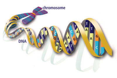
 A chromosome is a packaged unit of DNA and associated proteins.[1] The chromosome carries portions of the hereditary information of an organism. The DNA is tightly coiled many times around proteins called histones that support the chromosome structure.[2] The bound DNA units are called nucleosomes. Different kinds of organisms have different numbers of chromosomes. Humans have 23 pairs of chromosomes, 46 in all: 44 autosomes and two sex chromosomes. Each parent contributes one chromosome to each pair, so children get half of their chromosomes from their mothers and half from their fathers.
Contents
History[edit]
The word "chromosome" means "colored body" and comes from the Greek χρώμα or "chroma" for "color" and σομε or "some" for "body". By 1900 scientists knew that cells were the building blocks of living things. All living things start as a single fertilized cell which keeps dividing. Scientists identified tiny threads in the nucleus of the cells. Because they could stain the threads with colored dyes to study them under a microscope, they called the threads chromosomes, from the Greek words for colored bodies. They could see that chromosomes came in pairs, and that human cells all contained 23 matching pairs. American biologist Walter Sutton knew Mendel's principles of genetics work on peas, and suggested that chromosomes held the secret of inheritance.
Another American biologist, Thomas Hunt Morgan, developed the idea that chromosomes were made up of linked groups of factors called genes. He experimented with red-eyed fruit flies and found that sometimes a white-eyed fly appeared. When he mated them, he found that as he expected there were three red-eyed flies to every white-eyed fly, but that all the white-eyed flies were male. He concluded that the gene for white eyes must be on a chromosome that was related to being male. Later workers found that this is why some hereditary diseases such as hemophilia and muscular dystrophy only show in males, though women can carry the gene for the disease without showing it.
In 1991 a project called the Human Genome Project began to use computers to map the three billion base pairs which make up the 46 human chromosomes.
Physical Characteristics of Chromosomes[edit]
Most chromosomes have the rough shape of an I when they condense prior to replication and are as bits of long invisible string when unwound. The familiar X shape actually refers to 2 identical chromosomes referred to as sister chromatids. Each chromosome has a constriction point called the centromere, which divides the chromosome into two sections, or “arms.” The short arm of the chromosome is labeled the “p arm” for "petite". The long arm of the chromosome is labeled the “q arm” for "not petite". The location of the centromere on each chromosome gives the chromosome its characteristic shape, and can be used to help describe the location of specific genes.[3]
The chromosomal DNA molecule contains three specific nucleotide sequences which are required for replication: a DNA replication origin; the centromere to attach the DNA to the mitotic spindle.; a telomere located at each end of the linear chromosome.[4] There is also a telomere region within the human chromosome two, as well as a non-functional second centromere.[5] This indicates a historical chromosome fusion event.
Number of Chromosomes[edit]
As noted above, the chromosome number varies in different species. In humans there are 46 chromosomes, or 23 pairs of chromosomes (diploid), in every cell except the mature egg and sperm which have a set of 23 chromosomes (haploid).
Cell Division[edit]
Chromosomes are visible only during cell division, when the DNA is super coiled and condensed to facilitate distribution into daughter cells. Cell division in somatic cells (mitosis) results in the creation of daughter cells with the same number of chromosomes as the original cell, a total of 46 chromosomes in a human. Cell division in the germ cells, eggs and sperm (meiosis), results in the creation of daughter cells with half the number of chromosomes as the original cell (haploid cells). This reduction in the number of chromosomes is important so that the original number of chromosomes is restored following fertilization of the egg by the sperm.
Karyotype Analysis[edit]
The chromosome constitution of an individual, karyotype, can be analyzed following tissue culture of an appropriate sample. The most commonly used sample is blood (using the white blood cells or lymphocytes) since it is the most accessible. However, other samples are used depending upon the indication: amniotic fluid cells, to analyze the karyotype of the fetus; products of conception, to analyze the cause of a miscarriage or stillbirth; bone marrow cells, to diagnose the presence or type of leukemia; and skin, to determine the presence of another cell line (mosaicism).
Cell division is arrested during metaphase, when the chromosome material is condensed. Following hypotonic treatment and fixation, the cells are dropped on a slide and then stained. At least 20 metaphase spreads are analyzed and 2 or 3 metaphase spreads are photographed. The chromosomes are arranged from largest to smallest to create a karyotype.
Chromosomes vary in size and in shape. The pairs of autosomal chromosomes are arranged in a karyotype from the biggest to the smallest. The sex chromosomes are placed to the right of the smallest autosomes. Chromosomes vary in shape depending upon the position of the centromere, the structure that holds the two arms of the chromosomes together. If the centromere is in the middle, the chromosome is metacentric and the chromosome arms are equal in size. If the centromere is off center, the chromosome is submetacentric with a short arm labeled p (for petite) and a long arm labeled q (the next letter after p). If the centromere is close to the end, the chromosome is acrocentric and the very short arm consists of a stalk and a knob (satellite). Based upon size and shape, human chromosomes are divided into eight groups: A (1 to 3), B (4 and 5), C (6 to 12), D (13 to 15), E (16 to 18), F (19 and 20), G (21 and 22) and the sex chromosomes, XX in females and XY in males.
Chromosomes are further identified by banding patterns created by specialized staining procedures to produce G bands, R bands, C bands, etc. The choice of staining procedure depends upon the information that is desired. Most commonly, chromosomes are stained with trypsin-Giemsa to produce the G-banded pattern. The banding pattern is determined by the degree of chromatin condensation and the specific DNA-protein present at the different sites on the chromosome. These banding patterns are distinct and consistent for each chromosome. Thus, one can reliably identify the chromosome pairs (e.g., although of the same size and shape, the six acrocentric chromosomes in the D group can be differentiated into pairs 13, 14 or 15 based upon banding patterns). The bands are individually numbered (e.g., 11q23 refers to band #23 on the long arm q of chromosome #11). :[6]
References[edit]
- ↑ http://learn.genetics.utah.edu/units/disorders/karyotype/whatarechrom.cfm
- ↑ http://ghr.nlm.nih.gov/handbook/basics/chromosome
- ↑ http://www.rothamsted.ac.uk/notebook/courses/guide/chromo.htm
- ↑ http://ghr.nlm.nih.gov/ghr/chromosomes
- ↑ http://www.pnas.org/content/88/20/9051.abstract
- ↑ http://www.vivo.colostate.edu/hbooks/genetics/medgen/chromo/cytotech.html
Categories: [DNA] [Genetics]
↧ Download as ZWI file | Last modified: 03/10/2023 10:37:39 | 115 views
☰ Source: https://www.conservapedia.com/Chromosome | License: CC BY-SA 3.0
 ZWI signed:
 KSF
KSF