Epithelial, Endothelial
 From Britannica 11th Edition (1911)
 Epithelial, Endothelial and GLANDULAR TISSUES, in anatomy. Every surface of the body which may come into contact with foreign substances is covered with a protecting layer of cells closely bound to one another Epithelium. to form continuous sheets. These are epithelial cells (from θηλή, a nipple). By the formation of outgrowths or ingrowths from these surfaces further structures, consisting largely or entirely of cells directly derived from the surface epithelium, may be formed. In this way originate the central nervous system, the sensitive surfaces of the special sense organs, the glands, and the hairs, nails, &c. The epithelial cells possess typical microscopical characters which enable them to be readily distinguished from all others. Thus the cell outline is clearly marked, the nucleus large and spherical or ellipsoidal. The protoplasm of the cell is usually large in amount and often contains large numbers of granules.
The individual cells forming an epithelial membrane are classified according to their shape. Thus we find flattened, or squamous, cubical, columnar, irregular, ciliated or flagellated cells. Many of the membranes formed by Varieties. these cells are only one cell thick, as for instance is the case for the major part of the alimentary canal. In other instances the epithelial membrane may consist of a number of layers of cells, as in the case of the epidermis of the skin. Considering in the first place those membranes of which the cells are in a single layer we may distinguish the following:—
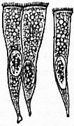
1. Columnar Epithelium (figs. 1 and 2).—This variety covers the main part of the intestinal tract, i.e. from the end of the oesophagus to the commencement of the rectum. It is also found lining the ducts of many glands. In a highly typical form it is found covering the villi of the small intestine (fig. 1). The external layer of the cell is commonly modified to form a thin membrane showing a number of very fine radially arranged lines, which are probably the expression of very minute tubular perforations through the membrane.
 |
 |
 |
| Fig. 1.—Isolated Epithelial Cells from the Small Intestine of the Frog. | Fig. 2.—Columnar Epithelial Cells resting upon a Basement Membrane. | Fig. 3.—Mosaic appearance of a Columnar Epithelial Surface as seen from above. |
The close apposition of these cells to form a closed membrane is well seen when a surface covered by them is examined from above (fig. 3). The surfaces of the cells are then seen to form a mosaic, each cell area having a polyhedral shape.
2. Cubical Epithelium.—This differs from the former in that the cells are less in height. It is found in many glands and ducts (e.g. the kidney), in the middle ear, choroid plexuses of the brain, &c.
 |
| Fig. 4.—Squamous Epithelial Cells from the Mucous Membrane of the Mouth. |
3. Squamous or Flattened Epithelium (fig. 4).—In this variety the cell is flattened, very thin and irregular in outline. It occurs as the covering epithelium of the alveoli of the lung, of the kidney glomerules and capsule, &c. The surface epithelial cells of a stratified epithelium are also of this type (fig. 4). Closely resembling these cells are those known as endothelial (see later).
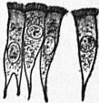 |
| Fig. 5.—Isolated ciliated Epithelial Cells from the Trachea. |
4. Ciliated Epithelium (fig. 5).—The surface cells of many epithelial membranes are often provided with a number of very fine protoplasmic processes or cilia. Most commonly the cells are columnar, but other shapes are also found. During life the cilia are always in movement, and set up a current tending to drive fluid or other material on the surface in one direction along the membrane or tube lined by such epithelium. It is found lining the trachea, bronchi, parts of the nasal cavities and the uterus, oviduct, vas deferens, epididymis, a portion of the renal tubule, &c.
In the instance of some cells there may be but a single process from the exposed surface of the cell, and then the process is usually of large size and length. It is then known as a flagellum. Such cells are common among the surface cells of many of the simple animal organisms.
When the cells of an epithelial surface are arranged several layers deep, we can again distinguish various types:—
 |
| Fig. 6.—A Stratified Epithelium from a Mucous Membrane. |
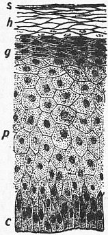 |
| Fig. 7.—Stratified Epithelium from the Skin. |
c, Columnar cells resting on the fibrous true skin. p, The so-called prickle cells. g, Stratum granulosum. h, Horny cells. s, Squamous horny cells. |
1. Stratified Epithelium (figs. 6 and 7).—This is found in the epithelium of the skin and of many mucous membranes (mouth, oesophagus, rectum, conjunctiva, vagina, &c.). Here the surface cells are very much flattened (squamous epithelium), those of the middle layer are polyhedral and those of the lowest layer are cubical or columnar. This type of epithelium is found covering surfaces commonly exposed to friction. The surface may be dry as in the skin, or moist, e.g. the mouth. The surface cells are constantly being rubbed off, and are then replaced by new cells growing up from below. Hence the deepest layer, that nearest the blood supply, is a formative layer, and in successive stages from this we can trace the gradual transformation of these protoplasmic cells into scaly cells, which no longer show any sign of being alive. In the moist mucous surfaces the number of cells forming the epithelial layer is usually much smaller than in a dry stratified epithelium.
2. Stratified Ciliated Epithelium.—In this variety the superficial cells are ciliated and columnar, between the bases of these are found fusiform cells and the lowest cells are cubical or pyramidal. This epithelium is found lining parts of the respiratory passages, the vas deferens and the epididymis.
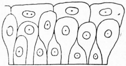 |
| Fig. 8.—Transitional Epithelium from the Urinary Bladder, showing the outlines of the cells only. |
3. Transitional Epithelium (fig. 8).—This variety of epithelium is found lining the bladder, and the appearance observed depends upon the contracted or distended state of the bladder from which the preparation was made. If the bladder was contracted the form seen in fig. 8 is obtained. The epithelium is in three or more layers, the superficial one being very characteristic. The cells are cubical and fit over the rounded ends of the cells of the next layer. These are pear-shaped, the points of the pear resting on the basement membrane. Between the bases of these cells lie those of the lowermost layer. These are irregularly columnar. If the bladder is distended before the preparation is made, the cells are then found stretched out transversely. This is especially the case with the surface cells, which may then become very flattened.
Considering epithelium from the point of view of function, it may be classified as protective, absorptive or secretory. It may produce special outgrowths for protective or ornamental purposes, such are hairs, nails, horns, &c., and for such purposes it may manufacture within itself chemical material best suited for that purpose, e.g. keratin; here the whole cell becomes modified. In other instances may be seen in the interior of the cells many chemical substances which indicate the nature of their work, e.g. fat droplets, granules of various kinds, protein, mucin, watery granules, glycogen, &c. In a typical absorbing cell granules of material being absorbed may be seen. A secreting cell of normal type forming specific substances stores these in its interior until wanted, e.g. fat as in sebaceous and mammary glands, ferment precursors (salivary, gastric glands, &c.), and various excretory substances, as in the renal epithelium.
Initially the epithelium cell might have all these functions, but later came specialization and therefore to most cells a specific work. Some of that work does not require the cell to be at the surface, while for other work this is indispensable, and hence when the surface becomes limited those of the former category are removed from the surface to the deeper parts. This is seen typically in secretory and excretory cells, which usually lie below the surface on to which they pour their secretions. If the secretion required at any one point is considerable, then the secreting cells are numerous in proportion and a typical gland is formed. The secretion is then conducted to the surface by a duct, and this duct is also lined with epithelium.
 |
| Fig. 9.—A Compound Tubular Gland. One of the pyloric glands of the stomach of the dog. |
Glandular Tissues.—Every gland is formed by an ingrowth from an epithelial surface. This ingrowth may from the beginning possess a tubular structure, but in other instances may start as a solid column of cells which subsequently Glands. becomes tubulated. As growth proceeds, the column of cells may divide or give off offshoots, in which case a compound gland is formed. In many glands the number of branches is limited, in others (salivary, pancreas) a very large structure is finally formed by repeated growth and subdivision. As a rule the branches do not unite with one another, but in one instance, the liver, this does occur when a reticulated compound gland is produced. In compound glands the more typical or secretory epithelium is found forming the terminal portion of each branch, and the uniting portions form ducts and are lined with a less modified type of epithelial cell.
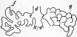
Glands are classified according to their shape. If the gland retains its shape as a tube throughout it is termed a tubular gland, simple tubular if there is no division (large intestine), compound tubular (fig. 9) if branching occurs (pyloric glands of stomach). In the simple tubular glands the gland may be coiled without losing its tubular form, e.g. in sweat glands. In the second main variety of gland the secretory portion is enlarged and the lumen variously increased in size. These are termed alveolar or saccular glands. They are again subdivided into simple or compound alveolar glands, as in the case of the tubular glands (fig. 10). A further complication in the case of the alveolar glands may occur in the form of still smaller saccular diverticuli growing out from the main sacculi (fig. 11). These are termed alveoli.
 |
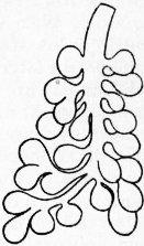 |
| Fig. 10.—A Tubulo-alveolar Gland. One of the mucous salivary glands of the dog. On the left the alveoli are unfolded to show their general arrangement. d, Small duct of gland subdividing into branches; e, f and g, terminal tubular alveoli of gland. | Fig. 11.—A Compound Alveolar Gland. One of the terminal lobules of the pancreas, showing the spherical form of the alveoli. |
The typical secretory cells of the glands are found lining the terminal portions of the ramifications and extend upwards to varying degrees. Thus in a typical acinous gland the cells are restricted to the final alveoli. The remaining tubes are to be considered mainly as ducts. In tubulo-alveolar glands the secreting epithelium lines the alveus as well as the terminal tubule.
The gland cells are all placed upon a basement membrane. In many instances this membrane is formed of very thin flattened cells, in other instances it is apparently a homogeneous membrane, and according to some observers is simply a modified part of the basal surface of the cell, while according to others it is a definite structure distinct from the epithelium.
In the secretory portion of the gland and in the smaller ducts the epithelial layer is one cell thick only. In the larger ducts there are two layers of cells, but even here the surface cell usually extends by a thinned-out stalk down to the basement membrane.
The detailed characters of the epithelium of the different glands of the body are given in separate articles (see Alimentary Canal, &c.). It will be sufficient here to give the more general characters possessed by these cells. They are cubical or conical cells with distinct oval nuclei and granular protoplasm. Within the protoplasm is accumulated a large number of spherical granules arranged in diverse manners in different cells. The granules vary much in size in different glands, and in chemical composition, but in all cases represent a store of material ready to be discharged from the cell as its secretion. Hence the general appearance of the cell is found to vary according to the previous degree of activity of the cell. If it has been at rest for some time the cell contains very many granules which swell it out and increase its size. The nucleus is then largely hidden by the granules. In the opposite condition, i.e. when the cell has been actively secreting, the protoplasm is much clearer, the nucleus obvious and the cell shrunken in size, all these changes being due to the extrusion of the granules.
Endothelium and Mesothelium.—Lining the blood vessels, lymph vessels and lymph spaces are found flattened cells apposed to one another by their edges to form an extremely thin membrane. These cells are developed from the Endothelium and mesothelium. middle embryonic layer and are termed endothelium. A very similar type of cells is also found, formed into a very thin continuous sheet, lining the body-cavity, i.e. pleural pericardial, and peritoneal cavities. These cells develop from that portion of the mesoderm known as the mesothelium, and are therefore frequently termed mesothelial, though by many they are also included as endothelial cells.
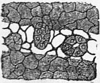 |
| Fig. 12.—Mesothelial Cells forming the Peritoneal Serous Membrane. Three stomata are seen surrounded by cubical cells. One of these is closed. The light band marks the position of a lymphatic. (After Klein.) |
A mesothelial cell is very flattened, thus resembling a squamous epithelial cell. It possesses a protoplasm with faint granules and an oval or round nucleus (fig. 12). The outline of the cell is irregularly polyhedral, and the borders may be finely serrated. The cells are united to one another by an intercellular cement substance which, however, is very scanty in amount, but can be made apparent by staining with silver nitrate when the appearance reproduced in the figure is seen. By being thus united together, the cells form a continuous layer. This layer is pierced by a number of small openings, known as stomata, which bring the cavity into direct communication with lymph spaces or vessels lying beneath the membrane. The stomata are surrounded by a special layer of cubical and granular cells. Through these stomata fluids and other materials present in the body-cavity can be removed into the lymph spaces.
Endothelial membranes (fig. 13) are quite similar in structure to mesothelial. They are usually elongated cells of irregular outline and serrated borders.
 |
| Fig. 13.—Endothelial Cells from the Interior of an Artery. |
By means of endothelial or mesothelial membranes the surfaces of the parts covered by them are rendered very smooth, so that movement over the surface is greatly facilitated. Thus the abdominal organs can glide easily over one another within the peritoneal cavity; the blood or lymph experiences the least amount of friction; or again the friction is reduced to a minimum between a tendon and its sheath or in the joint cavities. The cells forming these membranes also possess further physiological properties. Thus it is most probable that they play an active part in the blood capillaries in transmitting substances from the blood into the tissue spaces, or conversely in preventing the passage of materials from blood to tissue space or from tissue space to blood. Hence the fluid of the blood and that of the tissue space need not be of the same chemical composition.
↧ Download as ZWI file | Last modified: 11/17/2022 15:23:25 | 25 views
☰ Source: https://oldpedia.org/article/britannica11/Epithelial_Endothelial | License: Public domain in the USA. Project Gutenberg License
 ZWI signed:
 KSF
KSF